Article Text

Abstract
Background Evidence that mesenchymal stem cells (MSCs) derived from bone marrow, fat and umbilical cord can be used to treat refractory SLE and SLE nephritis is growing. MSCs were originally described as cells from bone marrow that have the capacity to differentiate into bone, cartilage and fat. More recently it has been recognised that all MSCs are peri-cytes and that their greatest potential is because they are pleiotropic and can both sense and repair their environment. We hypothesised that MSCs can reduce neutrophil activation in SLE by inhibiting neutrophil netting thus reducing induction of T-helper follicular cells that promote the development of long-living plasmablasts that can secrete autoantibodies.
Materials and methods We studied neutrophils derived from healthy donors and patients with SLE. Neutrophils were isolated using MACSxpress™ Neutrophil Isolation Kit (Miltenyi) and onto coverslips in 24-well plates and incubated for 1–2 hours with conditioned medium derived from MSCs or control medium. Netting was induced by culture ex vivo with 20 nM PMA for 2 hours. Coverslips were fixed in 4% paraformaldehyde and NETs were quantified using anti-human antibody directed against neutrophil elastase colocalizing with extracellular DNA using Hoechst 33342.
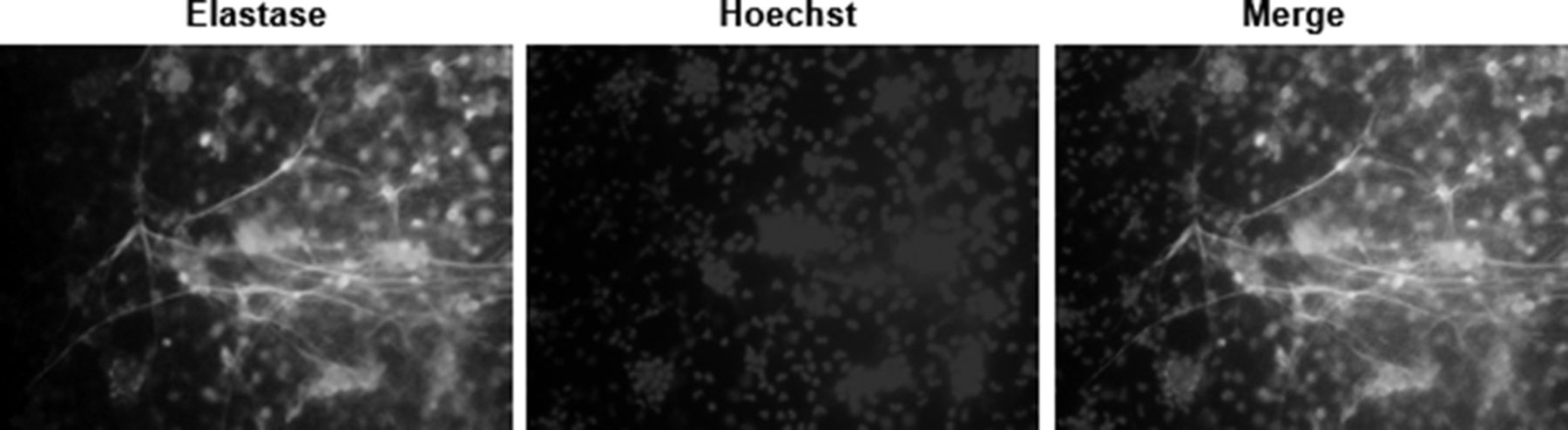
Results To date we have optimised the conditions of our assay. Studies are ongoing to determine the effect of MSCs and/or their products on neutrophil netting. Figure 1: seen below are netting neutrophils induced as described above. Assays are underway to determine the effect of MSCs on SLE netting neutrophils ex vivo.
Conclusion The possibility that MSCs and/or their products could act both on innate and adaptive immune responses in SLE is appealing. Demonstration of the effect of MSCs on neutrophils is critical in understanding the potential therapeutic role of MSCs in SLE and SLE related organ damage.

Neutrophil Elastase colocalized with extracellular DNA. Control neutrophils were isolated from peripheral blood and stimulated with 10nM PMA for 2 hr at 370C. Cells were fixed and stained for detection of neutrophil Elastase (green) and DNA was labeled with Hoechst 33342 (blue).
Acknowledgements This work has been supported by the Lupus Foundation of America and by the by the Clinical and Translational Science Collaborative of Cleveland, UL1TR000439 from the National Centre for Advancing Translational Sciences (NCATS) component of the National Institutes of Health and NIH roadmap for Medical Research. Its contents are solely the responsibility of the authors and do not necessarily represent the official views of the NIH.