Article Text

Abstract
Purpose Endothelial cell (EC) dysfunction is a hallmark of SLE and has been generally accepted to be one of the important factors contributing to the higher risk of thrombosis and atherosclerotic events observed in SLE patients. Although the presence of traditional factors (smoking, diabetes, increased age, obesity) and the presence of autoantibodies are associated with atherosclerosis and thrombotic events, they do not completely explain the higher risk of these events in SLE, suggesting the existence of other mechanism/factors.
Tie2 is a tyrosine kinase receptor essential for vascular development and blood vessel remodeling through interaction with its ligands angiopoietin-1 (Ang-1) and Ang-2. In homeostatic conditions, both Ang-1 and Ang-2 activate Tie2 signaling and induce vascular stabilization in a Tie1-dependent manner. However, inflammatory processes induce Tie1 cleavage, leading to the inhibition of Ang-1-induced Tie2 activation, and to the increase of Ang-2 now acting as a Tie2 antagonist, culminating in vascular dysfunction and EC activation 9–11. Importantly, this process has been implicated in both atherosclerosis and thrombosis.
As type I Interferons (IFN-α and IFN-β) are key cytokines in the pathogenesis of SLE, the aim of this study is to determine whether these cytokines induce Tie2 signalling-mediated endothelial cell dysfunction.
Methods Serum levels of Ang-1, Ang-2 and sTie1 in SLE patients (n=48) and healthy control (HC, n=29) were measured by ELISA. Human Umbilical Vein Endothelial Cells (HUVEC) were stimulated with IFN-α and IFN-β (both 1000 International Units –I.U.-) for 1, 2, 4, 6, 8, 12, 24, 48 and 72 hours. mRNA and protein expression of Ang-1, Ang-2, Tie1 and Tie2 were determined by quantitative PCR (qPCR) and ELISA, respectively. The phosphorylation of Tie2 determined by western blot and HUVEC viability was determined by calcein assay.
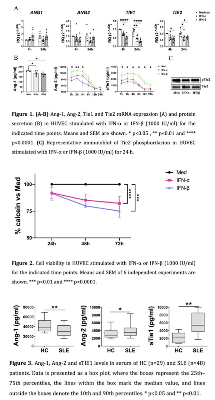
Results Type I IFNs, mainly IFN-β, significantly reduced the mRNA levels of TIE1 and TIE2. At level protein, IFN-β stimulation induced a significant increase in the secretion of the Tie1 ectodomain (sTie1). On the other hand, IFN-α and IFN-β did not modulate the mRNA expression of ANG1 or ANG2. However, both IFNs significantly reduced the protein secretion of Ang-1 after 24 h of stimulation. In the case of Ang-2, IFN-β induced Ang-2 secretion at early time points (<4h). Furthermore, IFN-α and IFN-β stimulation reduced Tie2 activation (Figure 1). At the functional level, both type I IFNs significantly reduced the viability of HUVEC (Figure 2).
Finally, and similarly to previous studies, we found reduced levels of Ang-1 and elevated levels Ang-2 in SLE patients compared to HC. Importantly, we showed for first time (to our knowledge) that sTie1 levels were also significantly elevated in SLE patients (Figure 3).

Conclusions Our results demonstrate that type I IFNs play a relevant role in the stability of endothelial cells by inhibiting Tie2 signaling, suggesting that these processes may be implicated in the cardiovascular events observed in SLE patients.
This is an open access article distributed in accordance with the Creative Commons Attribution Non Commercial (CC BY-NC 4.0) license, which permits others to distribute, remix, adapt, build upon this work non-commercially, and license their derivative works on different terms, provided the original work is properly cited, appropriate credit is given, any changes made indicated, and the use is non-commercial. See: http://creativecommons.org/licenses/by-nc/4.0/.