Article Text

Abstract
Purpose To identify whether IgG/IgM autoantibody ratios differ between patients with incomplete systemic lupus erythematosus (iSLE), patients with SLE and healthy controls (HC) and to assess whether IgG/IgM autoantibody ratios are related to progression from iSLE to SLE.
Methods In total, 34 iSLE patients, 41 SLE patients with quiescent disease and 22 HC were included in this cohort study. Patients were classified as iSLE if they met one clinical and one immunological criterion but less than four criteria of the Systemic Lupus International Collaborating Clinics (SLICC) criteria. Clinical assessment and blood collection was performed at baseline. Patients with iSLE received follow-up every six months up to five years or until progression to SLE. IgG and IgM levels for anti-dsDNA, anti-Ro52 and anti-Ro60 were measured by fluoro-enzyme-immuno-assay in serum samples obtained at baseline and follow-up. Optical density values were used for calculating the autoantibody ratios.
Results Cross-sectionally, anti-Ro52, anti-dsDNA and total IgG/IgM ratios did not differ between groups. Anti-Ro60 IgG/IgM ratios were significantly elevated in iSLE patients and SLE patients compared to HCs. There was no correlation between IgG/IgM ratios and interferon signature, SLEDAI score, complement levels or number of SLICC classification criteria at baseline. Of the 34 iSLE patients, six patients progressed to SLE and one patient developed primary Sjögren’s syndrome within five years. One patient that progressed to SLE showed an increase in anti-Ro52 and anti-Ro60 IgG/IgM ratio prior to progression. This increase was mainly due to an increase in anti-Ro52 and anti-Ro60 IgG autoantibodies. This finding was not observed in the other five progressors. IgG/IgM ratios were relatively constant during follow-up in non-progressors.

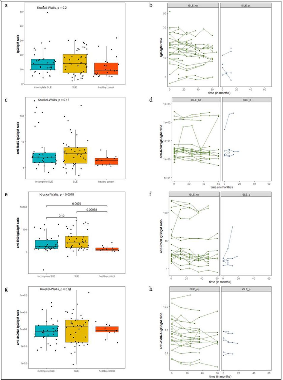
Median and interquartile ranges are depicted for a, c, e and g. Total IgG/IgM ratios (a), anti-Ro52 IgG/IgM ratios (c) and anti-dsDNA IgG/IgM ratios (g) were not significantly different between groups. Anti-Ro60 IgG/IgM ratios were significantly elevated in iSLE and SLE patients compared to healthy controls.Longitudinal line graphs of total IgG/IgM ratio (b), anti-Ro52 IgG/IgM ratio (d), anti-Ro60 IgG/IgM ratio (f) and anti-dsDNA IgG/IgM ratio (h) are shown for iSLE patients that did not progress to SLE (iSLE_np) and for iSLE patients that did progress to SLE (iSLE_p)
Conclusion Anti-Ro60 IgG/IgM ratios were significantly elevated in iSLE and SLE patients compared to HCs at baseline. IgG/IgM autoantibody ratios did not differ between iSLE patients that progressed to SLE and iSLE patients that did not progress.
This is an open access article distributed in accordance with the Creative Commons Attribution Non Commercial (CC BY-NC 4.0) license, which permits others to distribute, remix, adapt, build upon this work non-commercially, and license their derivative works on different terms, provided the original work is properly cited, appropriate credit is given, any changes made indicated, and the use is non-commercial. See: http://creativecommons.org/licenses/by-nc/4.0/.