Article Text

Abstract
An international summit on interferon (IFN) in inflammatory diseases, held in Gaithersburg, Maryland, USA (4–5 May 2017), united 22 internationally renowned clinicians and scientists with backgrounds in basic science, translational science and clinical medicine. The objectives of the summit were to assess the current knowledge of the role of type I IFN in inflammatory diseases and other conditions, discuss the available clinical trial data of anti-IFN therapeutic agents and identify key clinical and therapeutic knowledge gaps and future directions to advance the treatment landscape of diseases involving the type I IFN pathway. A discussion-based consensus process was used to assess three main clinical areas: the role of type I IFN in innate immunity, the role of type I IFN in autoimmune diseases and rational therapeutic targets in the IFN pathway. These are described here, along with current knowledge gaps and resulting recommendations. The advisors unanimously agreed that, despite significant obstacles, the field should transition from an organ-based model to a pathophysiology-based model. A better understanding of the molecular pathways could help inform potential therapeutic targets, thus progressing towards personalised medicine by tailoring the therapy to each patient.
- Interferon
- inflammatory diseases
- systemic lupus erythematous
This is an Open Access article distributed in accordance with the Creative Commons Attribution Non Commercial (CC BY-NC 4.0) license, which permits others to distribute, remix, adapt, build upon this work non-commercially, and license their derivative works on different terms, provided the original work is properly cited and the use is non-commercial. See: http://creativecommons.org/licenses/by-nc/4.0/
Statistics from Altmetric.com
Introduction
Type I interferon (IFN) is involved in the pathogenesis of systemic autoimmune diseases and several organ-targeted inflammatory diseases. Although its primary pathogenic role is well established for systemic lupus erythematosus (SLE), the role of type I IFNs in other autoimmune diseases is less clear. Evidence from analyses of peripheral blood of patients with rheumatoid arthritis demonstrating an IFN signature and evidence from murine models of diabetes mellitus strongly suggest a pathogenic role of type I IFN in a subset of these diseases.1 A more prominent type I IFN signature can be seen in Sjögrens syndrome, dermatomyositis (DM) and systemic sclerosis.2–5 Although we have gained a better understanding of the role of type I IFN in autoimmune and inflammatory diseases in recent years, key knowledge gaps remain, including those related to responses to distinct classes of therapeutic agents.
The inaugural International Summit on IFN in Inflammatory Diseases united 22 internationally renowned clinicians and scientists with backgrounds in basic science, translational science and clinical medicine. Goals of the summit included assessing the current knowledge of the role type I IFN plays in inflammatory diseases and other conditions, discussing available clinical data examining anti-IFN therapies and identifying current key clinical and therapeutic knowledge gaps. This report summarises the key findings of the meeting and includes the advisors’ recommendations for future efforts to advance the therapeutic landscape of diseases involving the type I IFN pathway.
Basic function of the type I IFN system
Role of IFN in immune responses
IFNs (types I–III) are a highly complex family of cytokines (type I IFN subtypes (IFN-α; 13 IFN-α genes, 12 IFN-α proteins in humans), IFN-β, IFN-ω, IFN-κ, and IFN-ε) with diverse effects on immune function. The type I IFN system serves as the major line of defence against viruses and microorganisms by promoting both innate and adaptive immune responses.6–8 The IFN concentrations required for antiviral and immunomodulatory effects may be distinct, and our understanding of how different type I IFNs and receptors generate diverse immune functions is still evolving.
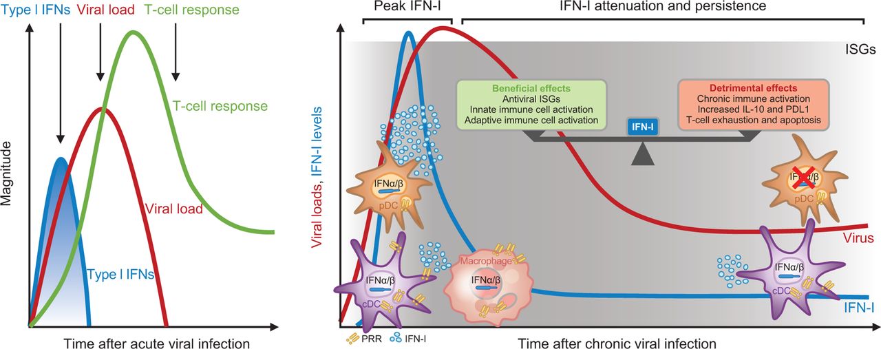
Type I IFNs are particularly important in antivirus host defence,6 and the complexity of the pathway may be necessary to defend against the variety of viruses that are encountered. The innate immune response to viral infection initiates intense but transient type I IFN expression (figure 1).9 10 Although we know many of the triggers that initiate a type I IFN response after an acute viral infection,8 the mechanisms by which this response is terminated are yet to be elucidated. The type I IFN pathway is dysregulated in SLE,11 12 and the advisors agreed that a large research gap exists in our understanding of what drives overproduction of this cytokine. Understanding the regulatory mechanisms that control the innate immune response may yield information beneficial to the autoimmune diseases community and may uncover novel drug targets.

The role of IFN in viral infection over time. cDC, conventional dendritic cell; IFN, interferon; IL, interleukin; ISG, interferon-stimulating genes; pDC, plasmacytoid dendritic cells; PD-L1, programmed death ligand 1; PRR, pattern recognition receptor. Left panel adapted from Crouse et al 9; right panel adapted from Zuniga et al 10 (Reprinted by permission from Springer Customer Service Centre GmbH: Springer Nature, Nature Reviews Immunology; Regulation of antiviral T cell responses by type I interferons. Crouse J, Kalinke U, Oxenius A, © 2015. Republished with permission of Annual Reviews, from Innate and Adaptive Immune Regulation During Chronic Viral Infections, Zuniga EI, Macal M, Lewis GM, et al, Vol. 2, © 2015; permission conveyed through Copyright Clearance Center.).
Although increased type I IFN expression is common in several diseases, the link between IFN signatures and various clinical manifestations of disease remains unclear, suggesting that the role of type I IFN may differ among diseases. The advisors discussed the potential utility of IFN blockade in HIV infection, because studies have demonstrated that IFN may be detrimental in these patients, potentially contributing to T-cell exhaustion.13 14 Many of the immune alterations that characterise patients with autoimmune diseases may be attributable to the immunomodulatory properties of type I IFN, epigenetic modifications and irreversible autoimmune processes.
Sources of IFN: plasmacytoid dendritic cells and other cells
Plasmacytoid dendritic cells (pDCs) have unique biology, are the most active type I IFN-producing cells and contribute to both innate and adaptive immunity (figure 2).15 16 pDC type I IFN production is most evident at early time points after viral infection. Over time, other cells (eg, conventional dendritic cells (cDCs) and epithelial cells) become more prominent producers of type I IFN.17–19 Exogenous (eg, viral) RNA and DNA elicit type I IFN production via toll-like receptor (TLR)–dependent and TLR-independent (cytosolic) mechanisms in pDCs and other cells.20 In autoimmune diseases, endogenous nucleic acids in the form of immune complexes can activate endosomal nucleic acid–sensing TLRs and drive type I IFN and IFN-induced gene expression, mimicking a viral infection.21

PDCs control many immune functions. APRIL, a proliferation-inducing ligand; BAFF, B cell activating factor; CCL, chemokine (C–C motif) ligand; ICOSL, inducible costimulator ligand; IDO, indoleamine dioxygenase; IFN, interferon; IL, interleukin; iNKT, invariant natural killer T cell; MHC, major histocompatibility complex; NK, natural killer; pDC, plasmacytoid dendritic cells; PD-L1, programmed death ligand 1; TGF, transforming growth factor; TH, T helper cell; TRAIL, tumour necrosis factor-related apoptosis-inducing ligand; TReg, T regulatory cell. Reproduced from Swiecki and Colonna16 (Reprinted by permission from Springer Customer Service Centre GmbH: Springer Nature, Nature Reviews Immunology; The multifaceted biology of plasmacytoid dendritic cells. Swiecki M, Colonna M, © 2015.).
Several nucleic acid sensors (eg, TLRs) within pDCs trigger distinct signalling pathways, including canonical type I IFN signalling—IFN-dependent activation of Janus kinase (JAK)–signal transducer and activator of transcription (STAT) leading to transcription of IFN-stimulated genes (the type I IFN gene signature (IFNGS)). Human pDCs express TLR7 and TLR9 nucleic acid sensors, and TLR7/TLR9 signalling induces a morphological change in pDCs, from an IFN-producing cell with high IFN production and low antigen presentation to an antigen-presenting cell with low IFN production and high antigen presentation.2 pDCs do not express TLR8 under most circumstances,22 which distinguishes them from monocytes, neutrophils and cDCs, although recent data from patients with systemic sclerosis suggest that TLR8 can be expressed in pDCs.23 Interleukin receptor–associated kinase-4 (IRAK-4) is involved in signalling innate immune responses from TLRs. IRAK-4-deficient patients produce IFN-α in response to viral infections but not to TLR7/TLR9 ligands; thus, these patients are susceptible to pyogenic bacterial infections (Gram+ bacteria) but appear to have normal resistance to viral infections.24 These data demonstrate the presence of non-TLR nucleic acid sensors.
Genetic contributors to the type I IFN system: SLE heterogeneity and interferonopathies
Understanding the molecular basis of diseases will help us progress towards personalised medicine by tailoring the therapy to the biological characteristics of each patient. Evidence suggests that genetic variations, along with varying exposure to environmental triggers and chance, may play a role in the heterogeneous manifestations of SLE.25 For example, SLE is more common in people with non-European-American ancestry26 and more severe in people with African-American ancestry.27 Serum IFN-α activity is greater in non-European patients with SLE than in Europeans, and the predominant genes associated with increased IFN concentrations differ between European-Americans and African-Americans.28 In addition, many genetic variants related to the IFN-α pathway are associated with SLE.29 Although we are still mapping the genetic associations with lupus in many populations, differences between populations clearly exist, and these variants affect immune phenotypes (eg, cytokine profile, autoantibodies). Evidence indicates that genetics is more strongly related to immune system phenotype than clinical phenotype, and the advisors suggested that future clinical trials should be stratified by immune phenotypes rather than clinical phenotypes. A combination of immune and genetic profiling is the future of assessing disease heterogeneity in lupus and should be considered in clinical trial design and potentially in patient management.
Type I interferonopathies result from mutations that lead to an overproduction of type I IFN and may be directly relevant to the pathology of lupus.30 Mutations similar to those found in interferonopathies have been identified in patients with lupus. Although most of these mutations are related to nucleic acid metabolism, other mutations associated with nucleic acid signalling, negative regulation of IFN production or response and proteasome function have been identified. These mutations primarily affect the brain, skin or both, and non-penetrance appears to contribute to variations in autoimmune phenotypes. Given that different mutations can have overlapping phenotypes, ultrasensitive assays of the cellular source and concentration of IFN may be useful to diagnose interferonopathies.31
IFN in autoimmune diseases
Disease mechanisms of lupus: interplay between IFN and immune cells
The interaction between type I IFNs and immune cells is complex.32 IFN promotes or primes the immune system and can be considered an immune system adjuvant. IFN-α induces B lymphocyte stimulator production, promoting B cell survival,33 and, conversely, B cells promote IFN-α production via pDCs.34 B cells have a dose-dependent effect on IFN-α production—as more B cells become activated, the production of type I IFN increases.34 In T cells, type I IFNs promote cell survival,35 and similarly to B cells, activated T cells enhance IFN-α production by generating some of the factors that recruit pDCs to produce IFN.36 Natural killer (NK) cells also stimulate IFN-α production, and the pDC–NK interaction enhances non-IFN cytokine production.37 38 Other cells have been implicated in type I IFN production. One study showed that when monocytes are added to NK-pDC cocultures, monocytes suppress the type I IFN response. For patients with SLE, this suppression may be reduced because of decreased production of reactive oxygen species.39 40 Thrombocytes have also been implicated in type I IFN production. Activated platelets can promote IFN-α secretion by pDCs.41
In SLE, the control of the type I IFN system is deficient, leading to an overproduction of IFN.42 Type I IFN appears to be a driver rather than a consequence of the disease, and type I IFN treatment is sufficient to elicit SLE-like diseases.43 44 Two pathways predominate as potential triggers for type I IFN activation in lupus: cell death or damage (eg, the lupus immune complex, oxidised DNA) and defective nucleic acid metabolism (eg, interferonopathies, intracellular nucleic acid accumulation). Although the origin of pathogenic nucleic acids in lupus is uncertain, potential sources include NETosis, apoptosis (in the context of inflammation), necroptosis, pyroptosis and autophagy.45 46 Increased generation or impaired degradation of cytosolic endogenous nucleic acids could activate cytosolic sensors. Oxidised DNA can be detected in skin lesions of patients with lupus and stimulates local type I IFN production.47 In the general population, nucleases protect from lupus development by degrading nucleic acids.48 Deoxyribonuclease 1 like 3 deficiency can manifest as lupus or an SLE-like disease.49 Autoantibodies to nucleic acids develop years before SLE (the type I IFNGS increases very early in the disease), and earlier detection may be possible in the future with more sensitive screening tools.
Influence of IFN on organ systems
Cardiovascular system
An imbalance between vascular damage and repair causes cardiovascular disease in patients with SLE. In these patients, IFN-α hampers vascular repair and inhibits angiogenesis,50–52 and the disease is an independent risk factor for development of atherosclerosis.53 Type I IFNs are independently associated with functional and anatomical markers of subclinical cardiovascular disease (ie, endothelial function, carotid plaque, severity of coronary calcification) in patients with SLE without a history of overt cardiovascular events, after controlling for Framingham risk factors.54
The prevalence of atherosclerosis has been found to be higher in patients with lupus compared with control individuals (37.1% vs 15.2%).55 Young women (35 and 44 years) with SLE have a 50-fold increased risk of vascular complications,56 including a greater prevalence of perioperative cardiovascular adverse events and greater mortality following first myocardial infarction, compared with patients without SLE.57 In atherosclerotic lupus-prone mice, IFN blockade improved vascular repair and decreased atherothrombosis.52 Inhibition of type I IFN receptor signalling via the JAK/STAT pathway with tofacitinib decreased vascular dysfunction in a murine lupus model.58
Central nervous system
Central nervous system (CNS) symptoms are present in approximately 40% of all patients with SLE and often occur in the first year of the disease.59 In some cases, symptoms may be associated with the presence of autoantibodies,60 but in most patients the underlying mechanisms are unknown. Current hypotheses around the pathophysiological mechanism of CNS symptoms in SLE involve increased production of peripheral IFN-α, which then enters the CNS and activates reactive microglia that engulf synaptic material, leading to reduced synaptic density in regions of the brain that affect behaviour.61 Repeated dosing with human IFN-α can promote a depression-like phenotype in mice similar to that observed in patients treated with recombinant IFN-α.62 63 In humans with lupus, a peripheral blood IFN signature is present in 90% of children and more than 50% of adults,64 and IFN-α is elevated in the cerebrospinal fluid of some patients.65
Muscle
Characterisation of the contribution of type I IFN to the immunopathogenesis of DM has relied on immunohistochemical analysis of muscle tissue along with gene expression studies. Although a previous view of DM pathogenesis focused on the deposition of autoantibodies in tissue and complement-mediated tissue damage, observations of expression of myxovirus resistance 1 protein, a type I IFN-induced gene product, in myofibres and capillaries pointed to a significant pathogenic role for type I IFN in DM.66 The muscle pathology of DM is unique, characterised by perifascicular atrophy. A similar topology of injury to keratinocytes and myofibres exists in the skin and muscle of patients with DM.67 DM gene expression mapping shows that induced genes are type I IFN regulated,68 69 and DM and SLE microarrays revealed type I IFN signalling far in excess of that of other skin diseases.70 Type I IFN-inducible proteins in muscle localise to areas of perifascicular atrophy as well as endothelial cells of capillaries and large vessels. A strong type I IFNGS is evident in the muscle, skin and blood of patients with DM.70–72
Joints
Joint involvement is common in lupus and is typically non-deforming and non-erosive. American College of Rheumatology criteria define lupus arthritis as characterised by tenderness, swelling or effusion in two or more joints, but many patients report joint pain in the absence of apparent synovitis. However, sensitive imaging (eg, with ultrasound) has revealed that subclinical synovitis is present in up to 40% of these patients.73
IFNs play a complex role in lupus arthritis. Expression of type I IFN-induced gene transcripts have been demonstrated in the synovial tissue of patients with SLE, and inhibition of osteoclastogenesis by IFN might account for the relatively non-erosive character of the arthritis seen in patients with lupus.74 Evidence of type I IFN activity in circulation has been detected in patients with lupus, and the anti-IFN-α receptor (IFNAR) therapeutic agent anifrolumab has been shown to reduce joint disease activity scores.75
Skin
Keratinocytes are a source of IFN-κ in healthy skin.76 IFN-κ maintains the basal IFN gene response in keratinocytes, induces the IFN gene response in fibroblasts and activates dendritic cells. IFNs promote a proinflammatory response in keratinocytes, including CXCL9, CXCL10 and interleukin-6 production.77 IFNs also have a role in protecting skin. In wild-type mice, ultraviolet (UV) light induces an IFN response that is independent of pDCs, requiring CCR2+ cells and may be protective against inflammation.78 Keratinocytes produce type I/II/III IFNs after viral infection and suppress viral replication.79
IFNs play a role in several skin diseases. In psoriasis, IFN-β is upregulated in skin lesions,80 although the exact role of IFNs remains unclear. UV light downregulates IFNAR1, which is thought to be relevant to the therapeutic efficacy of UV light for psoriasis.81 Subacute cutaneous lupus erythematosus is characterised by a robust type I IFNGS, which is correlated with disease activity.82 In these patients, IFN-κ production increases after UV exposure. A strong IFN signature is also present in skin lesions of patients with DM83 and Sjögren’s syndrome,84 and in interferonopathies, genetic mutations identify the importance of IFNs in skin disease.85
Rational therapeutic targets in the IFN pathway
Advisors agreed that a better understanding of the molecular pathways (eg, relative roles of TLR vs cytosolic nucleic acid sensors) could help uncover potential novel therapeutic targets,86 and future efforts should consider extracellular and intracellular drug targets upstream and downstream of IFN.87 88 Potential upstream candidates include cells producing IFN (eg, pDCs), nucleic acid receptors (eg, TLRs, cytosolic sensors), signalling components (eg, kinases, adaptors) and transcriptional regulators. Downstream candidates include IFNAR, signalling components (eg, JAK/STAT)89 and induced gene products (eg, IFN-stimulated genes, mRNAs, proteins). Therapeutic agents that target the type I IFN pathway (from pDC to JAK-STAT activation) in clinical development for IFN-driven diseases are listed in table 1. Although upstream drivers of type I IFN production may be more specific for SLE, they may differ for individuals, restricting efficacy to a subset of patients. For example, DNA-containing immune complexes may drive type I IFN production in some patients, whereas others may have immune complexes that predominantly contain RNA. The advisors also expressed concern that a greater risk of toxicity may exist when targeting signalling pathways downstream of the IFNAR because they are shared by many systems (eg, JAK/STAT signalling).
Therapeutic agents targeting components of the type I IFN pathway and in clinical development for IFN-driven diseases
Although complex cytokine/IFN signalling is an attractive focus, it can be a risky target (eg, the JAK2 knockout is embryonically lethal).90 Given there are four pairs of JAKs, JAK1–3 and TYK2, and JAKs exist as pairs on cytokine receptors, there will likely be a need for selective JAK inhibitors. Currently, two JAK inhibitors (ruxolitinib91 and tofacitinib92) are approved and are effective in allergic diseases. Tofacitinib is efficacious in murine lupus,58 and a Phase II clinical trial of baricitinib for the treatment of lupus has completed enrolment. Like all medications, JAK inhibitors can be associated with adverse events, including infection, cytopenias, increased lipids and gastrointestinal perforation, but are well tolerated by many patients.93
The advisors discussed the advantages and limitations of focusing on the type I IFN pathway as a therapeutic target. Given the uncertainty surrounding the roles of various upstream and downstream components of the IFN pathway in IFN-driven diseases, IFN represents a tractable drug target that is strongly linked to the IFNGS. Type I IFN plays an important role in the pathogenesis of SLE and likely in other disorders characterised by IFN signalling that have not been addressed previously. Anifrolumab is a fully human monoclonal antibody that binds to the type I IFN receptor and is in Phase III development for moderate to severe SLE. In the MUSE Phase IIb clinical trial,75 a high IFNGS was a biomarker of anifrolumab response compared with placebo. The outcomes of patients with a low IFNGS were not significantly different between those receiving drug and those receiving placebo. However, it is notable that the percentage of patients responding to the drug was roughly similar in the high and low IFNGS groups, and a relatively high placebo response was demonstrated in the low IFNGS group. The trial with anifrolumab supports the hypothesis that the type I IFN pathway is an important contributor to the pathophysiology of SLE. The data also suggest a need for additional research to understand whether there is a role for IFN pathway inhibition in therapy of those with a low IFNGS. Determining type I IFN pathway status in an individual patient based on an IFNGS test may help to predict which patients are more likely to respond to treatment.
Some advisors recognised several limitations of focusing on the type I IFN pathway as a therapeutic target, including safety concerns (eg, impaired antivirus protection, increased susceptibility to infections such as Herpes zoster), unclear long-term effects on the immune system, insufficient data on the link between high IFN and B-cell and plasma-cell activation and the potential alteration of the balance between IFN-α and IFN-β signalling in neurons if the antibody enters the CNS. In addition, given the diverse and complex alterations in many components of the immune response for patients with SLE and other autoimmune diseases, treatment with drugs that target the type I IFN pathway may not be sufficient to gain control of disease activity and may not treat all manifestations of the disease in a single patient; therefore, treatment would require several drugs.
Transitioning from an organ-based model to a pathophysiology-based model
The advisors unanimously agreed that, despite significant obstacles, the field should transition from an organ-based model to a pathophysiology-based model. Key obstacles to this approach include the necessity of finding biomarkers that identify the most relevant molecular pathways operative in a given patient; the challenges in establishing the clinically defined patient cohorts needed for investigation of the genetic variations and gene expression signatures associated with defined clinical features of disease and the uncertainty of categorising these disorders as IFN diseases, because other pathways also will be involved.
Based on these obstacles, the advisors recommended three types of pathophysiology-based studies. In the first type of study, a ‘basket approach’ design, two or three diseases with common phenotypes would be selected and specific outcomes would be measured. The basket approach would use an observational/open-label design with the composite endpoint based on the percentage of responders for each measure. In the second type of study, a ‘feature approach’ design, patients with a common feature (eg, arthritis) would be pooled and the response to treatment would be measured based on that common feature. The third type of study would use a ‘steroid-sparing approach’. Because many diseases are managed with steroids, a reduction in steroid use plus an additional clinical measure could be used as endpoints.
Future directions
After identifying and discussing knowledge gaps for the role of the type I IFN pathway in autoimmune diseases, the advisors generated a list of 13 key questions to be considered in future research efforts to advance the treatment landscape (table 2).
Key points to be considered in future research efforts
Conclusion
This summit was the first meeting in a series of meetings that unites clinicians and scientists with the goal of outlining future directions to explore novel treatment of diseases involving the IFN pathway. The advisors unanimously agreed that, despite significant obstacles, the field should transition from an organ-based model to a pathophysiology-based model. A better understanding of the molecular pathways could help inform potential therapeutic targets, progressing toward personalised medicine by tailoring the therapy to the characteristics of each patient.
Uncited online supplementary appendix 1.
Acknowledgments
The authors wish to thank the advisors for their contributions during the IFN Research Summit. Editorial support was provided by Lourdes W H Yun, MD, MSPH, and Francis Golder, BVSc, PhD, of JK Associates, as well as Michael A Nissen, ELS, of AstraZeneca.
References
- 1.↵
- 2.↵
- 3.↵
- 4.↵
- 5.↵
- 6.↵
- 7.↵
- 8.↵
- 9.↵
- 10.↵
- 11.↵
- 12.↵
- 13.↵
- 14.↵
- 15.↵
- 16.↵
- 17.↵
- 18.↵
- 19.↵
- 20.↵
- 21.↵
- 22.↵
- 23.↵
- 24.↵
- 25.↵
- 26.↵
- 27.↵
- 28.↵
- 29.↵
- 30.↵
- 31.↵
- 32.↵
- 33.↵
- 34.↵
- 35.↵
- 36.↵
- 37.↵
- 38.↵
- 39.↵
- 40.↵
- 41.↵
- 42.↵
- 43.↵
- 44.↵
- 45.↵
- 46.↵
- 47.↵
- 48.↵
- 49.↵
- 50.↵
- 51.↵
- 52.↵
- 53.↵
- 54.↵
- 55.↵
- 56.↵
- 57.↵
- 58.↵
- 59.↵
- 60.↵
- 61.↵
- 62.↵
- 63.↵
- 64.↵
- 65.↵
- 66.↵
- 67.↵
- 68.↵
- 69.↵
- 70.↵
- 71.↵
- 72.↵
- 73.↵
- 74.↵
- 75.↵
- 76.↵
- 77.↵
- 78.↵
- 79.↵
- 80.↵
- 81.↵
- 82.↵
- 83.↵
- 84.↵
- 85.↵
- 86.↵
- 87.↵
- 88.↵
- 89.↵
- 90.↵
- 91.↵
- 92.↵
- 93.↵
Footnotes
Contributors A list of meeting participants/advisors is provided in the appendix. MC and LR co-chaired the summit and contributed to manuscript preparation.
Funding This summit was fully sponsored by AstraZeneca, Gaithersburg, Maryland, USA.
Competing interests The two authors of this report, as well as the meeting participants, received fees for their time in preparing for and presenting at/attending this meeting. They received no fees for their work as authors of the manuscript.
Patient consent Not required.
Provenance and peer review Not commissioned; externally peer reviewed.