Article Text

Abstract
Patients with SLE display a significantly higher cardiovascular risk (CVR). Pulse wave velocity (PWV) has meanwhile been established as a reliable parameter of end-organ damage. Endothelial progenitor cells (EPCs) are critically involved in vascular repair under both physiological and pathological conditions. The aim of the study was to analyse PWV and the Vascular Augmentation Index (VAI) and EPC numbers/regeneration in a well-defined German SLE cohort. Thirty patients were included. Only two individuals displayed a PWV of above 10 m/s. There was no correlation between PWV percentiles and disease activity as reflected by the SLE Disease Activity Index. Neither EPC colonies nor percentages of circulating EPCs (CD133+/KDR+) correlated with PWV/VAI in a positive or negative manner. Thus, it can be questioned whether pulse wave analysis and/or EPC proliferation and circulating cell numbers are truly useful for CVR assessment in SLE.
- pulse wave velocity
- cardiovascular risk
- endothelial progenitor cells
This is an Open Access article distributed in accordance with the Creative Commons Attribution Non Commercial (CC BY-NC 4.0) license, which permits others to distribute, remix, adapt, build upon this work non-commercially, and license their derivative works on different terms, provided the original work is properly cited and the use is non-commercial. See: http://creativecommons.org/licenses/by-nc/4.0/
Statistics from Altmetric.com
Patients with SLE have a prevalence of cardiovascular disease up to 50 times higher than healthy individuals.1 Recently, we were able to show that regeneration of endothelial progenitor cells (EPCs) may be impaired in SLE.2 Aortic pulse wave velocity (PWV) measurement and the central Vascular Augmentation Index (VAI) serve as surrogate markers for arterial stiffness. They represent independent predictors of cardiovascular structural damage and correlate with clinical outcomes.3 ,4 The aim of this study was to correlate parameters of vascular stiffness with EPC regeneration/circulating EPCs in a well-defined German SLE cohort. The patients were consecutively recruited from the Clinic of Nephrology and Rheumatology of the University Hospital of Göttingen. Prevalences of the following comorbidities were arterial hypertension, 61%; smoking, 26%; use of steroids, 82% and statin therapy, 13% (table 1).
Baseline characteristics of patients
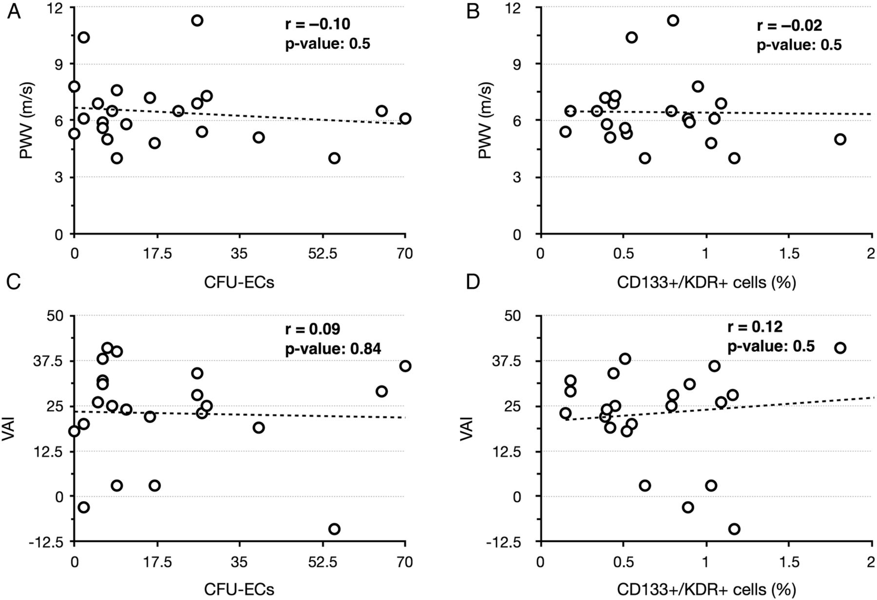
Analysis of PWV and VAI were successfully performed in 23 of 30 patients. All patients signed written consent to participate. The mean PWV was 6.4±1.7 m/s in all patients. Only two individuals displayed a PWV of above 10 m/s (10.4 and 11.3 m/s). PWV positively correlated with age (p=0.024). There was no correlation between PWV percentiles and disease activity as reflected by the SLE Disease Activity Index. The latter did also not correlate with the VAI percentiles, but anti-double stranded DNA levels correlated with the augmentation index in a negative manner (correlation coefficient −0.46, p=0.029). Neither EPC colonies nor percentages of circulating EPCs (CD133+/KDR+) correlated with PWV/VAI in a positive or negative manner (figure 1).

Correlation analysis between colony-forming unit-endothelial cells (CFU-ECs)/CD133+/KDR+ cells and pulse wave velocity (PWV)/vascular augmentation index (VAI). Neither cell regeneration, as reflected by the number of colonies growing in culture, nor percentages of circulating CD133+/KDR+ cells correlated with PWV or VAI in a positive or negative manner.
The most intriguing result of the current investigation was related to the absolute PWV values in patients with SLE with only two individuals displaying a PWV of above 10 m/s. A PWV of above 10 m/s has meanwhile been accepted as marker of cardiovascular end-organ damage.5 Castejon et al6 reported a mean PWV in SLE of 7.8±2.2 m/s with lower percentages of circulating EPCs in those patients displaying pathological PWV values. Nevertheless, the absolute range of PWV values in this particular category was not mentioned in the study. Comparable data have been reported by Valero-Gonzalez but once again, the absolute range of values is missing.7 A very early investigation was published in 2001, evaluating PWV in female patients with SLE with a mean PWV of 6.1±1.7 m/s.8 In a newer study, participants were subdivided into two subgroups (brachial-ankle PWV (baPWV) <14 vs >14 m/s). Nevertheless, the mean baPWV in all individuals was 15±0.38 m/s.9 Finally, Sacre et al10 reported a mean PWV of 6.3±0.8 m/s. Therefore, our observation has two implications: the absolute PWV may significantly vary in SLE, most likely depending on the subjects investigated and the method used for PWV analysis. In addition, EPC colony formation/circulating EPCs may not reliably correlate with parameters of vascular stiffness. Thus, it can be questioned whether pulse wave analysis and/or EPC proliferation and circulating cell numbers are truly useful for cardiovascular risk assessment in SLE. Finally, it needs however to be mentioned that the current study was performed in an uncontrolled manner which may account for some limitations.
Acknowledgments
Martha Potulski helped with initial studies.
Footnotes
Contributors PK wrote the manuscript, DP assisted in manuscript writing and analysed data, EH performed EPC analyses, TBN corrected the manuscript, GAM supplied financial support and SP designed the study and recruited all patients.
Funding This work was supported by the Heidenreich von Siebold Programm.
Competing interests None.
Patient consent Obtained.
Ethics approval The study was approved by the local ethics committee of the Georg August-University Göttingen. All participants signed written consent.
Provenance and peer review Not commissioned; externally peer reviewed.
Data sharing statement All data are presented in the manuscript.