Article Text

Abstract
Objective Patients with SLE have an increased risk of atherosclerosis (ATH) that is not adequately explained by traditional risk factors. We previously described the Predictors of Risk for Elevated Flares, Damage Progression, and Increased Cardiovascular disease in PaTients with SLE (PREDICTS) atherosclerosis-risk panel, which includes proinflammatory HDL (piHDL), leptin, soluble tumour necrosis factor-like weak inducer of apoptosis (sTWEAK) and homocysteine, as well as age and diabetes. A high PREDICTS score confers 28-fold increased odds for future atherosclerosis in SLE. The aim of this study is to determine whether PREDICTS biomarkers are modifiable by common lupus therapies.
Methods This prospective observational study included SLE subjects started on new lupus treatments. Leptin, sTWEAK, homocysteine and antioxidant function of HDL were measured at baseline (prior to drug initiation), 6 weeks and 12 weeks.
Results 16 subjects started mycophenolate (MMF), 18 azathioprine (AZA) and 25 hydroxychloroquine (HCQ). In MMF-treated subjects, HDL function progressively improved from 2.23 ± 1.32 at baseline to 1.37±0.81 at 6 weeks (p=0.02) and 0.93±0.54 at 12 weeks (p=0.009). sTWEAK levels also improved in MMF-treated subjects from 477.5±447.1 to 290.3±204.6 pg/mL after 12 weeks (p=0.04), but leptin and homocysteine levels were not significantly changed. In HCQ-treated subjects, only HDL function improved from 1.80±1.29 at baseline to 1.03±0.74 after 12 weeks (p=0.05). There were no changes in the AZA group. MMF treatment was still associated with significant improvements in HDL function after accounting for potential confounders such as total prednisone dose and changes in disease activity. Overall, the mean number of high-risk PREDICTS biomarkers at week 12 significantly decreased in the entire group of patients started on a new lupus therapy (2.1±0.9 to 1.8±0.9, p=0.02) and in the MMF-treated group (2.4±0.8 vs 1.8±0.9, p=0.003), but not in the AZA or HCQ groups. In multivariate analysis, the odds of having a high PREDICTS atherosclerosis risk score at 12 weeks were lower with MMF treatment (OR 0.002, 95% CI 0.000 to 0.55, p=0.03).
Conclusions 12 weeks of MMF therapy improves the overall PREDICTS atherosclerosis biomarker profile. Further studies will determine whether biomarker changes reflect decreases in future cardiovascular events.
- systemic lupus erythematosus
- atherosclerosis
- hydroxychloroquine
- mycophenolate
- azathioprine
This is an open access article distributed in accordance with the Creative Commons Attribution Non Commercial (CC BY-NC 4.0) license, which permits others to distribute, remix, adapt, build upon this work non-commercially, and license their derivative works on different terms, provided the original work is properly cited, appropriate credit is given, any changes made indicated, and the use is non-commercial. See: http://creativecommons.org/licenses/by-nc/4.0/.
Statistics from Altmetric.com
Introduction
There is a well-documented increased risk of atherosclerosis (ATH) in patients with SLE.1 Overall, there is a twofold to 10-fold increased risk of myocardial infarction in patients with SLE compared with the general population, with an even more striking 50-fold increased risk in younger women.2 Cardiovascular events may also result in greater morbidity and mortality in patients with SLE, as patients with SLE have higher risk of in-hospital mortality and prolonged length of hospitalisations compared with patients with diabetes and patients without SLE and diabetes.3 However, although there is an increase in traditional Framingham risk factors in patients with SLE, these traditional factors alone do not fully account for the increased risk of cardiovascular events.4 Thus, other novel inflammatory risk factors are likely to contribute to the increased ATH seen in SLE. Similar to the pathogenesis of other SLE disease manifestations, the formation of the atherosclerotic plaque is an inflammatory process, characterised by chronic oxidative damage, inflammatory lipid markers and immune cell activation. Identification of biomarkers that reflect the ongoing inflammation that underlies plaque formation will be critical for identifying therapeutic interventions that can halt or prevent this process.
Our group has previously identified several biomarkers that are associated with progression of carotid artery atherosclerosis in patients with SLE. For instance, we have shown that proinflammatory high-density lipoproteins (piHDL) are present more frequently in patients with SLE with carotid artery plaque than in those without plaque both in cross-section5 and longitudinally.6 Although HDL levels are traditionally protective against ATH morbidity and mortality, the relationship between HDL and ATH is complex and involves both the quantity and function of HDL.7 Traditional anti-inflammatory HDL has antioxidant properties; it removes reactive oxygen species (ROS) from low-density lipoproteins (LDL), protects LDL from oxidation and prevents subsequent recruitment of monocytes to the arterial wall.7 Proinflammatory HDL is unable to perform its usual protective role in the prevention of ATH. Although studies in the general population and in rheumatoid arthritis (RA) have shown that piHDL function improved with statin therapy (but not to normal levels),8 9 the impact of treatments commonly used for SLE on HDL function is unknown.
In addition to piHDL, our group has identified several other inflammatory biomarkers that are associated with plaque and intima-media thickness (IMT) progression in SLE. We recently discovered that when these biomarkers are combined into a panel, PREDICTS, a ‘high PREDICTS risk’ score confers 28-fold increased odds for carotid plaque in SLE women and is also associated with IMT progression. The biomarkers included are piHDL, soluble tumour necrosis factor (TNF)-like weak inducer of apoptosis (sTWEAK) (≥373 pg/mL), homocysteine ≥12 μmol/L, leptin ≥34 ng/dL, age ≥48 years and type 2 diabetes mellitus.6 Patients with three or more risk factors, or diabetes plus one additional risk factor, are considered to have ‘high’ PREDICTS risk. It is unknown, however, whether these biomarkers are modifiable by lupus disease-modifying agents.
We hypothesised that patients with SLE who are treated with disease-modifying treatments would have more favourable PREDICTS biomarker profiles, particularly in regard to piHDL, versus patients with SLE treated with other modalities.
Patients and methods
Subject selection
In this prospective observational study, we sequentially enrolled all patients with SLE in our cohort who were started on new lupus-modifying therapies in an 18-month period. Patients were excluded if any baseline SLE medication or statin was started or changed within the 12 weeks prior to study entry, or if any changes to background therapies were anticipated during the 12-week study period. If subjects had previously taken and discontinued a lupus therapy in the past and were restarted on this treatment again, they were allowed to enrol in the study as long as they had not taken the medication within the previous 6 months. Subjects were included in the analysis if they continued on medication for at least 6 weeks. All eligible participants fulfilled ≥4 of the 1997 revised American College of Rheumatology (ACR) criteria for classification as SLE.10 Although neither study participants nor treating physicians were blinded to study medication, biomarker assessments were performed in a blinded fashion. All participants provided written informed consent.
Data collection
Plasma samples were collected and cryopreserved at three time points: baseline (prior to initiation of drug), 6 weeks and 12 weeks postinitiation of therapy. On the day of plasma sampling, SLE disease activity was assessed using Safety of Estrogens in Lupus Erythematosus National Assessment-SLE Disease Activity Index (SELENA-SLEDAI).11 Plasma leptin (BioVendor, Candler, North Carolina, USA) and sTWEAK (R&D Systems, Minneapolis, Minnesota, USA) were measured using ELISA. HDL function was measured as described previously5 12 using a cell-free assay based on the ability of HDL to prevent oxidation. Normal HDL prevents oxidation of LDL and dichlorofluorescein diacetate (DCFH-DA), which releases a fluorochrome (DCF) on interaction with lipid oxidation products. To determine HDL function, the change in fluorescence intensity from oxidation of DCFH/LDL in the presence or absence of test HDL was measured. LDL was prepared from normal plasma as previously described,12 13 and HDL was prepared from test plasma using a dextran sulfate magnetic bead reagent.14 Twenty-five microlitres of LDL-C (100 µg/mL) was mixed with 6.25 µL of test HDL (100 µg HDL-C/mL) in black flat-bottom polystyrene microtitre plates and incubated at 37°C with rotation for 30 min. Twenty-five microlitres of 2.0 mg/mL DCFH solution was then added to each well, mixed and incubated at 37°C for 1 hour with rotation. Fluorescence was determined with a plate reader (Spectra Max, Gemini XS; Molecular Devices) at an excitation wavelength of 485 nm, emission wavelength of 530 nm and cut-off of 515 nm with photomultiplier sensitivity set at medium. Values of DCF activated by LDL in the absence of HDL were normalised to 1.0 fluorescence unit as the positive control. Values greater than 1.0 after the addition of test HDL indicated dysfunctional, piHDL; values less than 1.0 indicated anti-inflammatory (normal) HDL. In previously published studies, mean HDL function in healthy controls ranged from 0.44 to 0.66.8 15
Statistical analysis
Data were analysed using STATA V.14.0. Skewed continuous variables were logarithmically transformed to attain a normal distribution (note: non-transformed data are presented in figures and tables to facilitate interpretation of results). For variables that did not attain a normal distribution by logarithmic transformation, non-parametric tests were used. Study groups were compared at baseline using analysis of variance with Tukey’s analysis of individual columns. Changes in individual biomarker measurements over time were compared using paired samples t-test and the χ2 test or Fisher’s exact test for categorical variables. Either Pearson or Spearman rank correlation was calculated, dependent on if the variable was normally distributed. The significance level was set at p<0.05.
Multiple regression analysis was used to build models identifying risk factors associated with a change in PREDICTS biomarker status at 12 weeks. Generalised estimating equations (GEE) were performed to identify factors associated with changes in HDL function, sTWEAK and leptin over time and to account for the clustering of the data within subjects. Different covariance structures were examined, including independent, exchangeable, autoregressive of order 1 and unstructured matrices. Our statistical longitudinal models allowed for interaction terms between time and the treated groups so that each treated group could have different progression rates over the two time intervals. The quasi-likelihood based Akaike’s information criteria QIC and QIC_u criteria for generalised estimating equations were used to determine the best fitting models.16
Results
Ninety subjects were entered into the study: 20 subjects were prescribed mycophenolate mofetil (MMF), 22 subjects were prescribed azathioprine (AZA) and 27 subjects were prescribed hydroxychloroquine (HCQ). Twenty-one subjects were prescribed other therapies, including belimumab (n=5), methotrexate (n=4), cyclophosphamide (n=2), leflunomide (n=2), abatacept (n=1), tacrolimus (n=1), rituximab (n=1), and ciclosporin (n=1). Because of the small sample sizes in most of the treatment arms, data were only analysed for subjects started on MMF, AZA and HCQ. At week 6, 16 subjects were still taking MMF, 18 were taking AZA and 24 were taking HCQ; these subjects were included in the analysis. At the 12-week time point, 15 subjects were still taking MMF, 16 were taking AZA and 19 were still taking HCQ. Study subjects in all three groups had similar mean disease activity at study entry (p=ns), although patients in the MMF group were more likely to have active glomerulonephritis than patients in the HCQ group (p=0.05) and were also more likely to have a prior history of glomerulonephritis than subjects in either the AZA or HCQ groups (table 1).
Baseline demographic and clinical characteristics
HDL function is improved in patients with SLE taking MMF and HCQ, but not AZA
Overall, HDL function improved from baseline to 6 weeks (p=0.009) and from baseline to 12 weeks (p=0.001) in patients with SLE who were started on any new disease-modifying therapy (MMF, AZA or HCQ). We also examined HDL function changes in each individual treatment group. There were no statistically significant differences in baseline piHDL levels among the three treatment groups. In MMF-treated subjects, HDL function improved significantly from baseline after 6 weeks (p=0.02) and 12 weeks of therapy (p=0.009) (table 2). In HCQ-treated subjects, HDL function did not significantly change from baseline at 6 weeks of therapy; however, it did significantly improve after 12 weeks of therapy (p=0.05). In those treated with AZA, HDL function remained relatively stable at 6 and 12 weeks (p=ns).
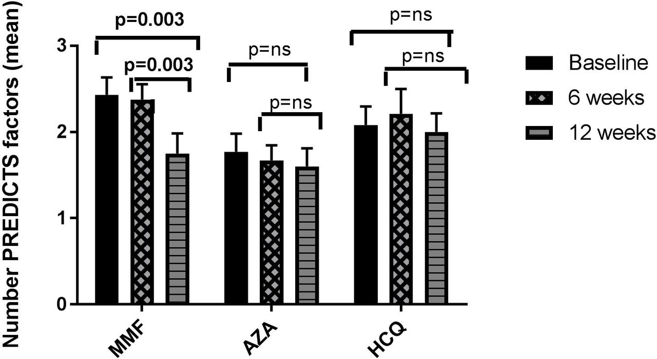
Changes in PREDICTS biomarkers over 12 weeks according to treatment subgroup
Improvement in HDL function is not dependent upon corticosteroid dose
The mean daily prednisone dose over the 12-week period was higher in the MMF-treated and AZA-treated groups than in the HCQ group (table 1). In order to account for the potential influence of prednisone dosage in the MMF, AZA and HCQ treatment groups, we divided each group into subjects taking high (≥10 mg/day) and low (<10 mg/day) daily prednisone doses. There were no significant differences in the percentage change of HDL function in high versus low prednisone groups in any of the treatment arms (data not shown). There were also no significant correlations between the mean daily prednisone dose or the total prednisone dose taken during the 12-week study period and per cent change of HDL function in the total cohort (p=ns) or in any individual treatment arm.
Improvement in HDL function is not dependent upon disease activity
There was no significant difference in disease activity at baseline among the three treatment groups. SELENA-SLEDAI did improve significantly by the 12-week time point in all three treatment groups. Although there was a strong correlation between per cent improvement in SLEDAI score and percent improvement in HDL function in the MMF group only (r=0.78, p=0.002), there was no significant correlation between changes in SLEDAI and changes in HDL function in the AZA or HCQ groups (figure 1).

Correlation between change in SLEDAI and change in pro-inflammatory HDL. The percent change in SLEDAI correlates with the per cent change in proinflammatory HDL from baseline to 12 weeks in (A) mycophenolate-treated patients, but not in (B) azathioprine-treated or (C) hydroxychloroquine-treated subjects. HDL, high-density lipoprotein; SLEDAI, SLE Disease Activity Index.
Improvement in HDL function in patients taking MMF is significant even after accounting for potential confounders
Generalised estimating equations were next used to examine the effects of three treatments (MMD, AZA and HCQ) on HDL function over three time points and adjusting for age, gender, ethnicity, SLEDAI and total prednisone use during the study period. After adjusting for these variables, patients in the MMF group exhibited a significant decrease in HDL function levels over time, with the estimated rates of decrease from baseline over the 12 weeks −0.71 (p=0.001). The difference in the progression rate for the AZA group and the rate in the MMF group was 0.63 (p=0.004), suggesting that AZA has a progression rate of about −0.08. The corresponding rate of decrease in the HCQ group was estimated to be −0.44 (p=0.31) and not statistically significant. This finding was consistent across different types of structures. All covariates, that is, age, gender, SLEDAI and total prednisone intake consumption have no significant effects on HDL function. This finding was consistent across different types of covariance structures. In addition, change in HDL function between baseline and at 6 weeks was significantly different in the MMF and AZA groups (p=0.012) and this significance difference persisted between baseline and at 12 weeks (p=0.005). All other covariates, that is, age, gender, SLEDAI and total prednisone consumption had no significant effects on HDL function. Among the different ethnic groups, only Asians’ HDL function responses differed significantly from that of Caucasians on average over time, but when ethnicity was grouped more broadly into Caucasians or non-Caucasians, this significance disappeared.
sTWEAK is improved in patients taking MMF but not AZA or HCQ
We next examined whether the other laboratory PREDICTS measurements changed in response to disease-modifying treatments. Neither leptin nor sTWEAK changed with the initiation of any new lupus therapy. Levels of sTWEAK did significantly decrease after 12 weeks of treatment with MMF (p=0.04) (table 2), but this difference was no longer significant in multivariate analysis. sTWEAK did not change in the AZA or HCQ treatment arms (table 2). Leptin levels did not significantly change over 12 weeks in any of the treatment arms.
The number of PREDICTS variables improved in MMF-treated patients
We also examined whether treatment with any disease-modifying therapy would result in a shift in biomarker values from a ‘high risk’ to a ‘lower risk’ PREDICTS category. We found that the addition of any new lupus therapy resulted in a decrease in the mean number of positive PREDICTS variables from 2.1±0.9 to 1.8±0.9 at 12 weeks (p=0.02). The percentage of patients with a high PREDICTS score, however, did not change significantly over 12 weeks (32% at baseline vs 33% at 12 weeks, p=ns). We also examined the impact of individual therapies on the overall PREDICTS score. We found that overall, the mean number of positive PREDICTS variables significantly decreased in the MMF-treated group from baseline to 12 weeks (p=0.03). There were no significant changes in the AZA or HCQ groups (figure 2).
The mean number of PREDICTS risk factors in each treatment group at baseline, 6 weeks and 12 weeks. PREDICTS scores range from a low of zero to a high score of 6. The biomarkers included are soluble tumour necrosis factor-like weak inducer of apoptosis (≥373 pg/mL), proinflammatory high-density lipoprotein, homocysteine ≥12 μmol/L, leptin ≥34 ng/dL, age ≥48 years and type 2 diabetes mellitus. The mean PREDICTS score decreased significantly from baseline in MMF-treated group over 12 weeks. AZA, azathioprine; HCQ, hydroxychloroquine; MMF, mycophenolate mofetil.
Logistic regression analysis determined which variables were associated with a high PREDICTS score at the 12-week follow-up. The model included baseline number of PREDICTS variables, lupus medications, total prednisone intake from 0 to 12 weeks and per cent change in SLEDAI over 12 weeks. Independent predictors for a high PREDICTS score at 12 weeks included the baseline number of PREDICTS variables (OR 20.6, 95% CI 3.3 to 128.6, p=0.001). Treatment with MMF was inversely associated with high PREDICTS at week 12, with an OR of 0.05 (95% CI 0.003 to 0.93, p=0.05) (see table 3).
Logistic regression for the association with a ‘high risk’ PREDICTS score* at 12-week follow-up
Discussion
In this prospective observational study, we found that patients with SLE who initiated treatment with a new disease-modifying therapy (MMF, AZA or HCQ) had improvements in both inflammatory HDL function and the number of high-risk PREDICTS variables. We also looked at the impact of different treatments on biomarkers of atherosclerosis and found that treatment with MMF for 12 weeks decreased two ‘high-risk’ biomarkers of atherosclerosis in patients with SLE: inflammatory HDL function (in both univariate and multivariate analysis) and sTWEAK (univariate analysis only). MMF therapy also resulted in a greater likelihood of improvement to a lower cardiovascular risk category using the PREDICTS model, but treatment with AZA did not. HCQ treatment did also result in improvements in HDL function in univariate analysis at 12 weeks, but no other significant changes to cardiovascular biomarkers were noted. Thus, treatment with MMF seems to be associated with better improvements in overall cardiovascular risk profile than other disease-modifying therapies tested. To our knowledge, this is the first study to demonstrate a change in novel biomarkers of atherosclerosis with disease-modifying therapies in patients with SLE.
There is some evidence to support MMF as an atheroprotective medication in SLE. MMF is a prodrug for mycophenolic acid (MPA), which inhibits inosine monophosphate dehydrogenase and decreases proliferating T and B cells.17 MPA also inhibits both lymphocytic and endothelial adhesion molecules, thereby decreasing lymphocyte infiltration into the atherosclerotic plaque.17 MPA also inhibits monocyte and macrophage recruitment to plaques.18 Thus, MMF may exert an atheroprotective effect in patients by altering the balance of inflammatory and protective arterial cell infiltration towards a more favourable phenotype. For example, in patients without SLE with carotid artery stenosis, 2 weeks of MMF therapy resulted in increased numbers of infiltrated regulatory T cells and decreased lesion-wide expression of inflammatory genes.19 In a separate study, MMF treatment in an ATH-prone SLE mouse model reduced atherosclerosis, recruitment of CD4+ T cells to atherosclerotic plaques and serum anti-oxidised LDL immunoglobulin G1 compared with statin treatment alone.20 In animal models of SLE and atherosclerosis, MMF treatment significantly reduced atherosclerotic burden in addition to reducing glomerulonephritis.20 21
MMF has several other potential antiatherogenic effects that may contribute to its ability to protect from ATH progression. MPA decreases oxidative stress and reduces formation of ROS by inhibiting interferon γ-stimulated and TNFα-stimulated inducible nitric oxide synthase (iNOS) activity.22 MMF also decreased other markers of oxidative stress in an animal model of cerebral ischaemia, including myeloperoxidase (MPO), glutathione, nitric oxide (NO) and malondialdehyde.23 In vitro, MMF attenuates MPO activity through inhibition of the toll-like receptor 4 (TLR4)/nuclear factor-kappa B signalling pathway.24 MMF also inhibits the activity of endothelial nicotinamide adenine dinucleotide phosphate oxidase oxidase (NOX), which in turn decreases endothelial superoxide formation and endothelial dysfunction.18 25 Given that MPO, NOX and NOS released during the process of NETosis have been implicated in the formation of dysfunctional piHDL,26 our findings that MMF therapy in patients with SLE improved HDL function should not be surprising.
There are scant published data regarding the atheroprotective effects of MMF therapy in lupus clinical studies. One subgroup analysis from a clinical trial of atorvastatin in patients with SLE did not find any reduction in measures of subclinical atherosclerosis in patients taking MMF; however, only 25 subjects were included in this analysis.27 There are some observational data supporting the atheroprotective effects of MMF therapy in the non-lupus population. In one study of renal transplant recipients, longer time on MMF was protective against aortic calcifications.28 MMF has also been shown to reduce intimal thickness in cardiac transplant patients compared with AZA-treated patients.29 Larger prospective randomised studies are needed to explore the impact of MMF treatment on cardiovascular disease in SLE.
Although several previous studies have demonstrated improvement in traditional lipid profiles with HCQ therapy,30–32 this is the first study to our knowledge that demonstrates improved HDL function in patients with SLE treated with HCQ. HCQ has also been shown to have other atheroprotective effects, including improved glycaemic control33 and reduced incidence of thrombotic events in SLE.34–37 Multiple retrospective cohort studies have demonstrated improved overall survival36 38 with HCQ use in SLE. HCQ has been associated retrospectively with decreased cardiovascular events in RA,39 and non-use of HCQ was associated with increased subclinical atherosclerosis in SLE.40 41 Although the exact mechanisms by which antimalarials exert protection are not well understood, one study in SLE-prone mice suggested that early treatment with HCQ prevents endothelial dysfunction via an antioxidant effect.42 A randomised controlled trial of HCQ versus placebo in preventing cardiovascular events in post-myocardial infarction (non-SLE) patients is currently underway.43 Larger prospective studies demonstrating a cardioprotective effect of HCQ in patients with SLE are needed.
In our study, therapy with AZA for 12 weeks failed to demonstrate any improvement in high-risk cardiovascular biomarkers. This is consistent with other studies in SLE that fail to demonstrate any cardioprotective effects with AZA. For example, AZA use was associated with increased cardiovascular events in the Spanish Registry of Systemic Lupus Erythematosus Patients of the Spanish Society of Rheumatology registry44 and in the multiethnic LUpus in MInoritypopulations: NAture versus Nurture cohort.45 AZA use was also associated with increased carotid IMT in the paediatric SLE Atherosclerosis Prevention in Pediatric Lupus Erythematosus cohort.46 Further studies will be needed to determine whether these associations are due to a direct effect of AZA or the inability of AZA to overcome the inflammation that leads to atherosclerosis.
Similar to our study, several studies have examined the impact of disease-modifying antirheumatic drugs (DMARDs) therapy on lipid levels and HDL function in RA. Although multiple investigations found that DMARD therapies in RA are associated with a paradoxical increase in hyperlipidaemia, there is some evidence to suggest that the increase in lipid levels is associated with improvement in HDL function. For instance, the Brigham Rheumatoid Arthritis Sequential Study registry study found that in patients with RA treated with DMARDs and/or anti-TNF agents, improvement in C-reactive protein was associated with increased LDL function and improvement in HDL function (measured as cholesterol-efflux capacity).47 Tocilizumab therapy in patients with RA also altered HDL composition towards a more anti-inflammatory phenotype, despite increases in LDL concentrations.48 The net impacts of lipid changes on cardiovascular risk in RA are not fully understood, however, and further longitudinal studies will be required in both RA and SLE to clarify the role of DMARDs in the management of cardiovascular risk.
There are several limitations to our study. First, this was an observational study, so the subjects were not randomised to a treatment group. In addition, background medication use was not dictated by the study protocol. Patients in the MMF and AZA treatment groups did have significantly higher prednisone use than those in the HCQ group. Even after accounting for cumulative prednisone dose during the 12-week period in multivariate analysis, however, MMF was still associated with improvements in HDL function and with the total PREDICTS score. Sixty-three per cent and 83% of subjects in the MMF and AZA groups, respectively, were also taking background HCQ, so it is difficult to determine any additive impact of HCQ on cardiovascular biomarkers. However, all subjects had been on stable doses of HCQ (and any other non-steroid lupus therapies) for at least 12 weeks prior to initiation, and no changes to HCQ dose were allowed during the investigational period. It is also possible that patients in the different medication groups might have had differing levels of compliance with therapy, which could decrease the effect sizes seen. Finally, although it is encouraging that cardiovascular biomarker changes were observed during the 12-week duration of this observational study, longer studies will be necessary to determine whether improvements in cardiovascular risk profiles with MMF and HCQ are sustained over time. Further studies will also be required to determine if therapeutic lowering of cardiovascular risk markers reduces future cardiovascular events.
In conclusion, HDL function is significantly improved over 12 weeks in patients with SLE treated with MMF or HCQ, although not to normal levels. There was no significant improvement in piHDL function in patients treated with AZA over 12 weeks, despite significant improvements in SELENA-SLEDAI scores. Using logistic regression, only MMF was significantly associated with improvement of PREDICTS cardiovascular risk profile. Therefore, MMF might offer not only control of glomerulonephritis in some patients but might also provide some protection from accelerated atherosclerosis in patients with SLE.
References
Footnotes
Contributors All authors were involved in drafting the article or revising it critically for important intellectual content and approved the final version to be published. MM had full access to all of the data in the study and takes responsibility for the integrity of the data and the accuracy of the data analysis. MM, JG, BHH and BJS contributed to the study conception and design. MM, JG, BHH, WC, BJS and LS contributed to the acquisition of data. MM, WKW and BJS contributed to the analysis and interpretation of data.
Funding Initial work was funded by an investigator- initiatedgrant from Aspreva pharmaceuticals. Further work funded by grants from National Institute of Arthritis and Musculoskeletal and Skin Diseases at the National Institute of Health R01AR063754-01A1(to MM), and K01AR059095 (to BS).
Competing interests MM has received honoraria from Astra Zeneca and Glaxo Smith Klein. BHH has received grant funding from Janssen Pharmaceuticals and Bristol Myers Squibb. All other authors have declared no conflicts of interest.
Patient consent for publication Not required.
Ethics approval The University of California Institutional Review Board approved the study protocol (#07-02-025-02).
Provenance and peer review Not commissioned; externally peer reviewed.
Data availability statement Data are available upon request.