Article Text

Abstract
Objective Recurrent disease flare is one of the key problems in lupus patients. A Chinese Flare-Prevention Lupus Initiative Cohort (FLIC) was established. Risk factors of disease flare were evaluated accordingly.
Methods Patients with low-grade disease activity (the Safety of Estrogens in Lupus Erythematosus National Assessment–SLE Disease Activity Index (SELENA-SLEDAI) =≤6, daily prednisone ≤20 mg, no British Isles Lupus Assessment Group A or no more than one B organ domain score) from January 2014 to August 2020 were included in the FLIC. Disease flares were defined by the modified SELENA-SLEDAI Flare Index. Low disease activity status (LDAS) and remission were also assessed. The cumulative flare rate was estimated by an event per 100 person-years analysis. Cox proportional hazards models were performed to identify risk factors of subsequent disease flares after adjusting clinical confounders. Survival was assessed with the Kaplan-Meier method.
Results 448 eligible patients with low-grade disease activity were included in FLIC. During a mean follow-up of 30.4 months, 170 patients flared. The cumulative lupus flare rate was 22.2 events per 100 patient-years. Compared with patients without flare, those with lupus flares were taking more prednisone, had higher disease activity index and with less patients attained LDAS/remission at baseline. They also had higher rates of antiphospholipid antibodies (aPLs) and antiribosomal P antibody. Cox regression analysis confirmed that attainment of either LDAS or remission at baseline were independent protective factors against subsequent disease flare (LDAS but not in remission: HR 0.58, 95% CI 0.38~0.88; remission: HR 0.46, 95% CI 0.30~0.69), while aPL was a risk factor of lupus flares (HR 1.95, 95% CI 1.36~2.78). Kaplan-Meier curves indicated that attaining LDAS or remission and absence of aPL at baseline had the least flare risk.
Conclusions In our real-world cohort study, not attaining LDAS or remission at baseline and aPL positivity was associated with higher risk of disease flares in patients with low-grade SLE.
- Systemic Lupus Erythematosus
- Antibodies, Antiphospholipid
- Therapeutics
Data availability statement
Data are available on reasonable request. Researchers will need to send requests to the corresponding authors with protocols to gain access.
This is an open access article distributed in accordance with the Creative Commons Attribution Non Commercial (CC BY-NC 4.0) license, which permits others to distribute, remix, adapt, build upon this work non-commercially, and license their derivative works on different terms, provided the original work is properly cited, appropriate credit is given, any changes made indicated, and the use is non-commercial. See: http://creativecommons.org/licenses/by-nc/4.0/.
Statistics from Altmetric.com
WHAT IS ALREADY KNOWN ON THIS TOPIC
Recurrent disease flare followed by organ damage is a key problem in lupus patients with low-grade disease activity.
WHAT THIS STUDY ADDS
The single-centre Chinese Flare-Prevention Lupus Initiative Cohort first aimed at identifying risk factors of lupus flares.
Our real-world data indicated that the presence of antiphospholipid antibodies (aPLs) and not attaining low disease activity status or remission at baseline were risk factors of subsequent lupus flares.
HOW THIS STUDY MIGHT AFFECT RESEARCH, PRACTICE AND/OR POLICY
Our data reinforce the importance of treat to target in lupus management and identified aPLs as another risk factors for lupus flares. Our data pave the way towards patient-tailored flare prevention strategies in the future.
Introduction
SLE is a chronic systemic autoimmune disease with flare–remission pattern as its typical feature.1 Even for patients with low-grade disease activity, recurrent lupus flare followed by organ damage is still a key problem that remains unsolved.1 2 As for active lupus patients, most of current clinical trials3–6 are aiming to reduce disease activity indexes as primary endpoints, such as SLE Responder Index7 or British Isles Lupus Assessment Group (BILAG) based Combined Lupus Assessment.8 However, keeping lupus in quiescence and preventing disease relapse is also an important outcome measurement for relative stable lupus patients.2 9 10 There were studies concerning the determinants of disease flares in general SLE population.11 12 However, in the real-world, understanding the frequency and risk factors of subsequent lupus flares for patients with low-grade disease activity is still incomplete.
Therefore, a Chinese Flare-Prevention Lupus Initiative Cohort (FLIC) was constructed, and the current study is attempting to address this question in this population.
Methods
We conducted this real-world study in a single centre, Shanghai Renji Hospital, South Campus. All the consecutive patients with low-grade disease activity during June 2014 and August 2020 meeting the following inclusion criteria were included in the study: (1) fulfilled the 1997 revised American College of Rheumatology criteria for SLE13; (2) scores of the Safety of Estrogens in Lupus Erythematosus National Assessment–SLE Disease Activity Index (SELENA-SLEDAI)14 ≤ 6 at screening,14 and with no BILAG15 A or no more than one B organ domain score; (3) received a stable treatment regimen with fixed doses of prednisone (0–20 mg/day), and/or hydroxychloroquine (HCQ), and/or immunosuppressive agents (IS) for at least 30 days. (4) Patients’ informed consent was obtained, and patients were admitted/discharged within 1 day in the day-care centre of our institution with a systemic evaluation as the baseline. Patients were arranged to subsequent outpatient follow-up and a yearly systemic check-up as aforementioned.
Two treat-to-target (T2T) criteria were assessed. Low disease activity status (LDAS) was defined as SLEDAI ≤4, with prednisone dose ≤7.5 mg/day; no activity in any major organ or no new disease activity features; HCQ and IS were allowed. Remission was described as clinical SLEDAI score=0, with prednisone ≤5 mg/day, HCQ and IS as maintenance.16
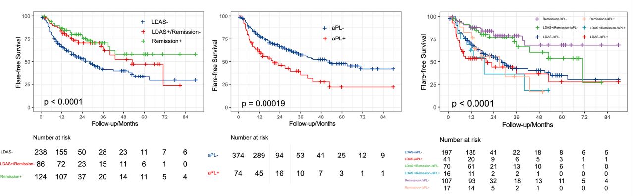
Three subgroups in FLIC were assessed, patients with more active disease not fulfilling LDAS at baseline (LDAS−); patients attained LDAS but not in remission (LDAS+/remission−); patients in remission at baseline (remission+).
Disease flares including major flares and mild-to-moderate flares were defined according to the modified SELENA-SLEDAI Flare Index (SFI)17 18 without the physician global assessment (PGA) item. The intention-to-treat item was used to determine the severity of disease flares, that is, for mild-to-moderate flares, prednisone dose was increased but ≤0.5 mg/kg/day or HCQ or non-steroidal anti-inflammatory drugs (NSAIDs) was added; for major flares, the prednisone dose was increased to >0.5 mg/kg/day or an escalation of IS treatment.
Detection of extractable nuclear antigen antibodies panel were performed by immunoblotting assay. Antiphospholipid antibodies (aPLs) include anticardiolipin, anti-β2-glycoprotein 1 antibody, both IgG and IgM, which were detected by ELISA and lupus anticoagulant assessed according to the international guidelines.19 Anti-ds-DNA antibody was also monitored by ELISA.
The clinical data were expressed as the n (%) or mean±SD. Continuous parameters were compared with the Mann-Whitney U test, and categorical parameters were analysed by Fisher’s exact test. The cumulative flare rate was estimated by an event per 100 person-years analysis. Cox proportional hazards analysis was applied to identify risk factors of disease flares adjusting confounders identified by univariate difference along with experts’ opinions based on clinical significance. Flare-free survival was assessed with the Kaplan-Meier method.
All statistical analyses were conducted in the Graphpad V.5.0 or SPSS V.22.0 software package. P<0.05 was considered statistically significant.
Results
During this period, among 1198 consecutive lupus patients presented to our centre, the cohort of FLIC with 448 eligible patients at low-grade disease activity was established (figure 1). A percentage of 91.3 (n=409) of these patients were female, with an average age of 34.3 years. They had a baseline mean SLEDAI of 2.47±2.00 and a daily prednisone dose of 8.59±4.98 mg. A percentage of 89.1% and 59.6% of them were taking HCQ and IS, respectively. At the time of enrolment, 210 patients (46.9%) attained LDAS, of which 124 were in remission. Other baseline characteristics including serological status were listed in table 1.
Baseline characteristics of 448 lupus patients with low-grade disease activity
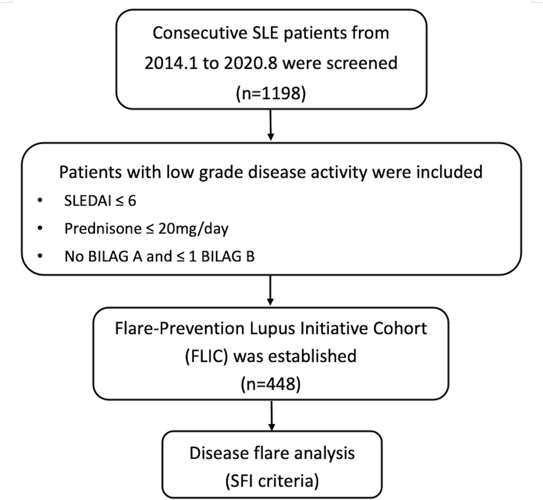
Flow chart of this study. SFI, SELENA-SLEDAI Flare Index
During a mean follow-up of 30.4 months, 170 patients had disease flares. Of these flare events, 55.3% (n=94) were manifested as major flares including 31 new-onset or relapsing lupus nephritis, 12 refractory rash, 11 thrombocytopaenia, 10 neuropsychiatric manifestations, 9 constitutional symptoms, 7 severe arthritis and muscular involvement, 5 mesenteric vasculitis, 3 serositis, 2 pulmonary arterial hypertension, 1 myocarditis, 1 macrophage activating syndrome, 1 autoimmune hepatitis and 1 hyperimmunoglobulinaemia. The patient with hyperimmunoglobulinaemia had a strikingly elevated polyclonal IgG up to 39.5 g/L and an erythrocyte sedimentation rate up to 114 mm/hour over time. The treating physician decided to initiate rituximab which, by SFI definition, is a significant escalation of treatment; therefore, a major flare event was recorded. The cumulative lupus flare rate and major flare rate were 22.2 and 12.7 events per 100 patient-years, respectively. The mean duration of first lupus flare since entry was 13.39±13.04 months.
Compared with patients who did not flared, those with flare events had higher positivity of aPL (flare: 24.7% vs no flare: 11.5%, p=0.014) and antiribosomal P antibody (flare: 25.9% vs no flare: 16.2%, p=0.01). The patients with flare had lower rate of baseline LDAS (34.1% vs 54.7%, p=<0.001) and remission attainments (17.6% vs 33.8%, p=0.0002), along with higher SLEDAI (2.9±1.9 vs 2.2±2.0, p=0.0004) and higher daily prednisone dose (9.99±4.80 vs 7.74±4.90 mg, p<0.0001). Demographic characters and HCQ usage were comparable between two groups (table 2).
Baseline characteristics of patients who had or had no disease flares
In order to identify risk factors of lupus flares in SLE patients with low-grade disease activity, Cox logistic regression analysis was performed. Variables with univariate difference along with experts’ opinions based on clinical significance were chosen. Eight candidate parameters including gender, age, disease duration, aPL, baseline attainment of LDAS/remission, usage of HCQ and exposure of IS entered into regression model. It was confirmed that aPL was an independent risk factor of subsequent disease flares with the HR of 1.95 (95% CI 1.36~2.78), while attaining LDAS or remission at baseline were protective factors against flares (LDAS+/remission−: HR 0.58, 95% CI 0.38~0.88; remission+: HR 0.46, 95% CI 0.30~0.69) (table 3). Furthermore, attainment of LDAS/remission (LDAS+/remission−: HR 0.48, 95% CI 0.27~0.84; remission+: HR 0.35, 95% CI 0.20~0.62) and the presence of aPL (HR 2.13, 95% CI 1.36~3.32) were also protective/risk factors in the regard of major flares.
Risk factors of subsequent disease flares in patients with low-grade disease activity by Cox regression analysis
Kaplan-Meier curves demonstrated that patients who were in remission or LDAS at entry and were negative for aPL had a higher flare-free survival (figure 2).

Flare-free survivals of patients with or without risk factors. (A) With or without APL; (B) with or without attaining LDAS/remission at baseline; (C) with or without two factors. LDAS, low disease activity status.
Discussion
SLE is a chronic autoimmune disease. It has been reported that SLE follows three different courses: long-term quiescent, relapsing–remitting and persistently active. The relapsing–remitting pattern is the most frequent clinical type.20 In the real world, there are large unmet needs for those patients with low-grade disease yet at risk of subsequent disease relapses.2 Reports concerning the prevalence and risk factors of lupus flares in this population were still insufficient. To the best of our knowledge, FLIC is the first cohort enrolling SLE patients with low-grade disease activity and is aimed at investigating preventive strategies for lupus flares.
In our FLIC, the cumulative flare rate was 22.2 events per 100 patient-years during a median follow-up of 30.4 months, with a baseline mean SLEDAI of 2.47±2.00 and a daily prednisone dose of 8.59±4.98 mg. Independent risk factors including aPL and not attaining LDAS or remission at baseline were identified. Of note, in this study, we applied the simplified definitions of LDAS and remission due to the lack of PGA data as compared with the original LLDAS criteria21 and DORIS remission criteria.22 However, the simplified definitions without PGA has been used in studies and validated in multiethnic lupus cohorts.16 23 24 It has been reported that T2T strategy using those definitions as endpoints was associated with less flares,21 25 and our results confirmed this concept.
The flare rates of lupus patients varied between different studies, populations and cohorts.3 12 26 27 Focusing on the specific group of patients with low-grade disease, the flare rate is roughly in parallel with the baseline disease activity status. For instances, the flare rate of lupus patients in remission was reported as around 5%–27% per patient-year.11 28 As comparison, according to the data of our previous metformin lupus flare prevention trial,9 the flare rate in the placebo arm was 35.5 events per 100 patient-years. The flare rate in FLIC is in between, probably due to a higher LDAS attainment at entry (46.9%) than that in the metformin trial (38.6%).24 Our data underscored that remission or LDAS attainment at baseline incurred a twofold reduction of subsequent flare risk (HR: 0.46–0.58) among SLE patients with low-grade disease.
As to another risk factor, it had been demonstrated that either positive for aPL or confirmed diagnosis of APS was related with lupus flares during pregnancy.29 30 However, there was no universal agreement about whether it could increase the risk of disease flare in general. In our Chinese FLIC population with low-grade disease, the presence of aPL turned out to be an independent risk factor, especially combining with not attaining LDAS/remission at baseline. Furthermore, the prevalence of aPL (16.5%) seemed to be numerically lower than previous reports in Chinese SLE cohorts including ours (20%–30%).31–35 The meaning and underlying cause is unknown.
It is noteworthy that discontinuation or reduction of HCQ was associated with lupus flares.36 However, in our cohort, the association of disease flare with baseline HCQ usage was not observed. It might because of the majority of patients in FLIC (~90%) received HCQ, which left the number of non-HCQ patients too small to make a difference.
There were several limitations in this study. First, this was a single-centre study with relatively moderate sample size and limited follow-up time. Second, accrual of damage was not evaluated. Third, no predefined protocol for de-escalation of therapy might serve as an important confounder that contributes to disease flare. The expansion of the study in the aforementioned directions is warranted. Nevertheless, our data pave the way towards patient-tailored flare prevention strategies in patients with SLE in the future.
Data availability statement
Data are available on reasonable request. Researchers will need to send requests to the corresponding authors with protocols to gain access.
Ethics statements
Patient consent for publication
Ethics approval
The protocol of this study complied with the recommendations of the Declaration of Helsinki and was approved by the ethics committees of Renji Hospital.
References
Footnotes
FS and LZ contributed equally.
Contributors All authors took part in revising the article and approved the final version to be published. SY and TL contributed to the conception and design, and were guarantors of the study. HW, DZ, JC and XW were responsible for collecting the data. FS and LZ performed the analysis and drafted the article.
Funding SY reports a grant from Clinical Research Plan of SHDC (No. SHDC2020CR1015B); FS reports grants from Shanghai Municipal Health Commission (No. 202040291).
Competing interests None declared.
Provenance and peer review Not commissioned; externally peer reviewed.