Article Text

Abstract
Objective Studies show that generic cardiovascular risk (CVR) prediction tools may underestimate CVR in SLE. We examined, for the first time to our knowledge, whether generic and disease-adapted CVR scores may predict subclinical atherosclerosis progression in SLE.
Methods We included all eligible patients with SLE without a history of cardiovascular events or diabetes mellitus, who had a 3-year carotid and femoral ultrasound follow-up examination. Five generic (Systematic Coronary Risk Evaluation (SCORE), Framingham Risk Score (FRS), Pooled Cohort Risk Equation, Globorisk, Prospective Cardiovascular Münster) and three ‘SLE-adapted’ CVR scores (modified Systematic Coronary Risk Evaluation (mSCORE), modified Framingham Risk Score (mFRS), QRESEARCH Risk Estimator V.3 (QRISK3)) were calculated at baseline. The performance of CVR scores to predict atherosclerosis progression (defined as new atherosclerotic plaque development) was tested with Brier Score (BS), area under the receiver operating characteristic curve (AUROC) and Matthews correlation coefficient (MCC), while rank correlation was tested with Harrell’s c-index. Binary logistic regression was also applied to examine determinants of subclinical atherosclerosis progression.
Results Twenty-six (21%) of 124 included patients (90% female, mean age 44.4±11.7 years) developed new atherosclerotic plaques after a mean of 39.7±3.8 months’ follow-up period. Performance analysis showed that plaque progression was better predicted by the mFRS (BS 0.14, AUROC 0.80, MCC 0.22) and QRISK3 (BS 0.16, AUROC 0.75, MCC 0.25). c-Index showed no superiority for discrimination between mFRS and QRISK3. In the multivariate analysis, QRISK3 (OR 4.24, 95% CI 1.30 to 13.78, p=0.016) among the CVR prediction scores and age (OR 1.13, 95% CI 1.06 to 1.21, p<0.001), cumulative glucocorticoid dose (OR 1.04, 95% CI 1.01 to 1.07, p=0.010) and antiphospholipid antibodies (OR 3.66, 95% CI 1.24 to 10.80, p=0.019) among disease-related CVR factors were independently associated with plaque progression.
Conclusions Application of SLE-adapted CVR scores such as QRISK3 or mFRS, as well as monitoring for glucocorticoid exposure and the presence of antiphospholipid antibodies, can help to improve CVR assessment and management in SLE.
- systemic lupus erythematosus
- atherosclerosis
- ultrasonography
- cardiovascular diseases
Data availability statement
Data are available upon reasonable request. All data analysed in this study are available by a reasonable request to the corresponding author.
This is an open access article distributed in accordance with the Creative Commons Attribution Non Commercial (CC BY-NC 4.0) license, which permits others to distribute, remix, adapt, build upon this work non-commercially, and license their derivative works on different terms, provided the original work is properly cited, appropriate credit is given, any changes made indicated, and the use is non-commercial. See: http://creativecommons.org/licenses/by-nc/4.0/.
Statistics from Altmetric.com
WHAT IS ALREADY KNOWN ON THIS TOPIC
Despite accumulating data on the increased cardiovascular disease risk (CVR) in SLE compared with the general population, evidence about the performance of traditional (generic) CVR assessment tools versus those incorporating SLE diagnosis (SLE-adapted) is inconclusive. Subclinical atherosclerosis is frequent in SLE and predicts future cardiovascular events. This study examines for the first time the performance of multiple generic and ‘SLE-adapted’ cardiovascular risk (CVR) scores to predict vascular ultrasound-detected atherosclerosis progression in SLE.
WHAT THIS STUDY ADDS
SLE-adapted scores such as the modified Framingham Risk Score and QRESEARCH Risk Estimator V.3 (QRISK3) performed better than generic risk tools to predict atherosclerotic plaque progression in SLE. QRISK3 among CVR scores, and cumulative glucocorticoid exposure and antiphospholipid antibodies among disease-related risk factors were independently associated with new plaque development.
HOW THIS STUDY MIGHT AFFECT RESEARCH, PRACTICE OR POLICY
Generic risk scores compared with SLE-adapted tools are less efficient to predict the progression of subclinical atherosclerosis in SLE, which may lead to suboptimal management of CVR. Application of SLE-adapted risk scores, as well as strict monitoring of disease-specific features such as glucocorticoid exposure and antiphospholipid antibody (aPL) positivity, may improve CVR assessment and management in SLE. Validation of our findings in larger studies will help to improve cardiovascular health in these patients.
Introduction
Cardiovascular disease is one of the most common causes of death, along with infections, in SLE,1 2 a prototype rheumatic and musculoskeletal disorder (RMD) primarily affecting young adults. Cardiovascular risk (CVR) in patients with SLE is 2-fold to 10-fold higher compared with the general population,3 4 with higher relative risk of myocardial infarction and stroke reported in women of younger age.5–7 Subclinical atherosclerosis, an early indicator of cardiovascular disease,8 is also frequent in patients with SLE with a threefold to fourfold increased risk of carotid plaques compared with the general population9–11 and a higher or comparable risk versus other diseases of increased CVR burden, such as rheumatoid arthritis and diabetes mellitus (DM).12
Measures for CVR assessment in SLE include generic CVR prediction tools and ‘SLE-adapted’ risk equations that incorporate the presence of SLE and/or disease features.13 14 The validity of various generic CVR prediction tools is not fully elucidated, while evidence about the preferential use of disease-specific risk tools is inconclusive.13–19 The recent ‘EULAR Recommendations for CVR Management in RMDs Including SLE and Antiphospholipid Syndrome’ did not endorse the use of any particular CVR assessment tool due to limitations of the current evidence.12
Aside from the established role of generic risk calculators to predict long-term cardiovascular events in the general population,8 some studies have also shown their predictive value to assess atherosclerosis progression on vascular imaging.20–22 Due to high atherosclerosis burden and the high risk of asymptomatic plaque progression in SLE associated with future cardiovascular events,8–11 the evaluation of multiple generic and SLE-adapted scores as potential predictors of subclinical atherosclerosis progression in patients with SLE is of high clinical significance.
The aim of this study is to examine whether various generic and SLE-modified CVR scores can predict the development of new atherosclerotic plaques in a 3-year follow-up vascular ultrasound study in SLE.
Methods
Study population
We included all eligible patients followed up in our academic centre who fulfilled the 2012 Systemic Lupus International Collaborating Clinics classification criteria for SLE23 and had a vascular ultrasound examination at baseline and after 3 years of follow-up by the end of March 2020 (COVID-19 outbreak in Greece). Patients with SLE who concurrently fulfilled the criteria for antiphospholipid syndrome (APS) were also included.24 We excluded from data analysis patients with a history of clinically established coronary artery disease (angina, myocardial infarction or coronary artery revascularisation procedure) or cerebrovascular disease (stroke, transient ischaemic attack or carotid artery revascularisation procedure) of atherosclerotic origin or atherosclerotic peripheral artery disease and patients with DM. Eight patients declined the follow-up ultrasound examination (flow chart, online supplemental figure S1).
Supplemental material
Study design
All study participants underwent a baseline vascular ultrasound assessment for the detection of atherosclerotic plaques in the carotid and femoral arteries. The participants were re-evaluated by vascular ultrasound approximately 3 years after the baseline visit. The following baseline characteristics were recorded at the time of baseline and the 3-year follow-up vascular ultrasound: age, disease-related features (disease duration, disease activity assessed by the Systemic Lupus Erythematosus Disease Activity Index 2000 (SLEDAI-2K), disease damage assessed by the Systemic Lupus International Collaborating Clinics/American College of Rheumatology Damage Index (SLICC/ACR DI), antinuclear and anti-dsDNA antibodies, C3 and C4 levels, antiphospholipid antibodies including anticardiolipin and anti-β2GPI antibodies and lupus anticoagulant, which were measured and considered positive according to the laboratory classification criteria for APS,24 lupus nephritis and chronic kidney disease (defined as the presence of glomerular filtration rate <60 mL/min), and medications: immunosuppressives, glucocorticoids use and their cumulative dose at the end of follow-up and during the 3-year follow-up period, while hydroxychloroquine, antihypertensives, statins and antiplatelets were recorded at baseline, at the end of follow-up, and during the follow-up period (reported as use over the 75% and 100% of time between the baseline and last follow-up visit). CVR factors included history of smoking (current or ever, pack-years), arterial hypertension (defined as use of antihypertensive treatment or blood pressure higher than 139/89 mm Hg as average of three sequential readings with 1 min interval in the supine position after at least 10 min of rest), hyperlipidaemia (defined as use of lipid-lowering medication and/or total cholesterol ≥240 mg/dL and/or low-density lipoprotein ≥130 mg/dL and/or triglycerides ≥160 mg/dL), obesity (body mass index ≥30 kg/m2) and family history of coronary artery disease.
Vascular ultrasound examination
All vascular ultrasound evaluations were performed by the same blinded assessor (GK). Subclinical atherosclerosis was assessed by the presence of plaques in a total of eight arterial beds (left and right, common and internal, carotid arteries and carotid bulb, and both common femoral arteries) by ultrasound. Atherosclerotic plaques were defined as the local increase of the intima–media thickness (IMT) of >50% compared with the surrounding vessel wall, an IMT of >1.5 mm or local thickening of >0.5 mm.25 Progression of atherosclerosis was defined as either (1) new appearance of plaques in individuals without plaques at baseline or (2) increase in number of plaques in individuals who already had plaques at the same carotid or femoral artery. Measurements were performed using high-resolution B-mode ultrasound (Vivid 7 Pro, GE Healthcare) with a 12 MHz linear matrix array transducer.
CVR scores
The following five generic CVR scores were calculated for all study participants: the Systematic Coronary Risk Evaluation (SCORE) score26; the Framingham 10-year risk score for coronary heart disease (Framingham Risk Score (FRS))27; the Pooled Cohort Risk Equation (PCRE) score28; the Globorisk score29; and the Prospective Cardiovascular Münster (PROCAM) score.30 Additionally, three SLE-adapted scores were calculated for all participants: the modified Systematic Coronary Risk Evaluation (mSCORE) (a 1.5-times multiplier to SCORE is applied),8 the modified Framingham Risk Score (mFRS) (a 2.0-times multiplier to FRS is applied)31 and the QRESEARCH Risk Estimator V.3 (QRISK3), which includes SLE as an independent CVR factor.32 Details about each score are presented in online supplemental table 1.
Statistical analysis
All data were assessed for normal distribution. Categorical variables were presented as frequencies and percentages and compared with the χ2 test. Continuous variables were presented as mean±SD or median with IQR; subgroup comparison was made with Student’s t-test or Mann-Whitney U test, as appropriate. The performance of each of eight CVR scores to predict the progression of subclinical atherosclerosis was assessed with Brier Score (BS), Matthews correlation coefficient (MCC) and receiver operating characteristic (ROC) curves, while rank correlation was tested with Harrell’s c-index.
Logistic regression models were used to assess determinants of atherosclerotic plaque progression. Demographic, clinical, laboratory and treatment parameters (including baseline and end of follow-up medication use, use for at least 75% of the follow-up period or the entire follow-up period (100%), and cumulative dose (in gram) for corticosteroids) were included in the univariate analysis and those found statistically significant (p<0.1), along with clinical considerations, were entered in the multivariate model. We additionally performed a second model (model 2) including only the variables with a p value of <0.05 in the univariate analysis. At a second step, one generic or SLE-adapted risk score was added each time in the logistic regression model to examine their potential role as independent predictors for plaque progression. A cut-off value of p<0.05 was set to denote statistical significance except for the univariate analysis where the cut-off p value was <0.1. Statistical analysis was performed using SPSS V.25.0 and MedCalc analysis V.14.2.
Results
In total, 124 Caucasian patients with SLE (90% female, mean age 44.4±11.7 years, mean disease duration 8.8±7.5 years) were included in the study. The mean follow-up period was 39.7±3.8 months, and the range of the time span between the two ultrasound examinations was 30–47 months. The mean SLEDAI-2K was 2.4±3.0, and the SLICC/ACR DI was 0.8±1.0. Thirty-eight patients fulfilled also the classification criteria for APS.24
At baseline, atherosclerotic plaques were evident in 30 patients, and more specifically carotid plaques were detected in 25 patients, femoral plaques in 19, and at both sites in 14 patients. After a mean of 39.7±3.8 months’ follow-up, progression of subclinical atherosclerosis (new atherosclerotic plaques) was observed in 26 patients: 9 with and 17 without the presence of plaques at baseline. Characteristics of study participants and risk estimates by the different generic and SLE-adapted CVR scores at baseline are shown in tables 1 and 2, respectively.
Demographics and clinical characteristics at baseline of 124 patients with SLE with or without vascular ultrasound-confirmed atherosclerosis and with or without progression of atherosclerosis
Mean values of generic and SLE-adapted cardiovascular risk scores at baseline of 124 patients with SLE with or without vascular ultrasound-confirmed atherosclerosis and with or without progression of atherosclerosis
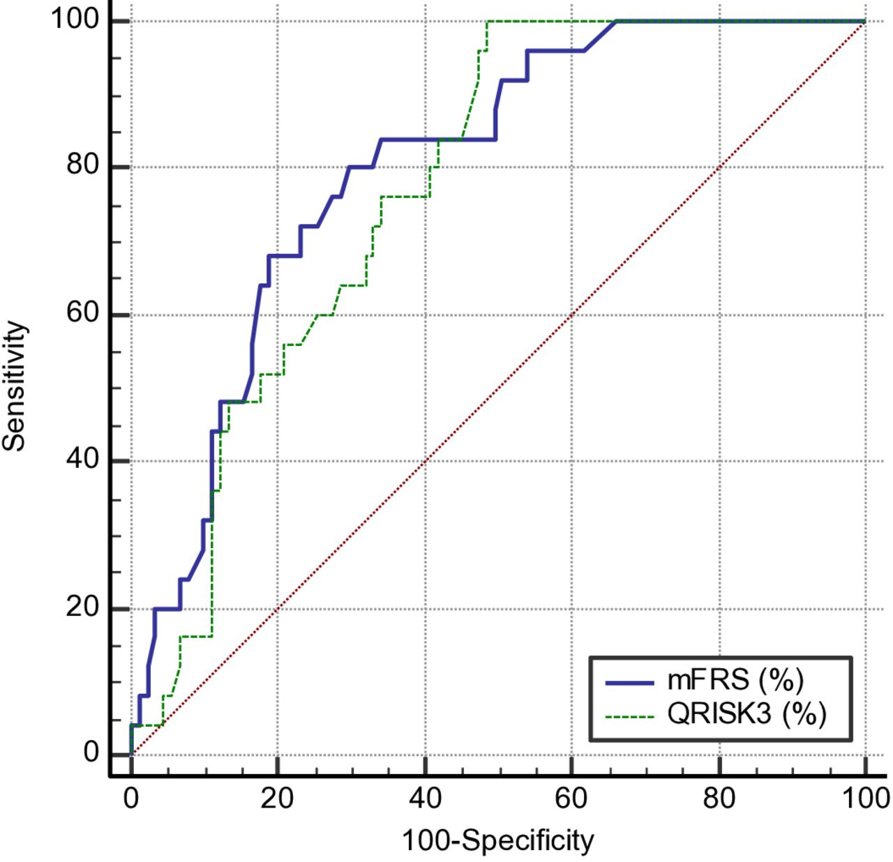
The performance of five generic and three SLE-adapted CVR scores to predict the progression of subclinical atherosclerosis, according to BS, ROC analysis and MCC, is depicted in table 3 and figure 1 and in online supplemental file 1. As presented, mFRS and QRISK3 showed the best overall performance (BS 0.14 and 0.16, respectively) and classification ability (MCC 0.22 and 0.25, respectively). Additionally, ROC analysis displayed a fair-to-good discriminatory capacity for all risk models (area under the curve (AUC) values from 0.66 to 0.80), while both mFRS and QRISK3 presented the highest AUC among examined scores. The c-index showed no superiority for discrimination between mFRS and QRISK3 (figure 1).
Performance of cardiovascular risk prediction models for plaque progression in 124 patients with SLE
Receiver operating characteristic analysis evaluating the performance of mFRS and QRISK3 to predict the progression of subclinical atherosclerosis in patients with SLE (discriminatory difference in area under the curve compared by c-index: 0.80 vs 0.75, p=0.37). mFRS, modified Framingham Risk Score; QRISK3: QRESARCH Risk Estimator V.3.
Multivariate analysis of plaque progression showed that age (OR 1.13, 95% CI 1.06 to 1.21, p<0.001), cumulative glucocorticoid dose (OR 1.04, 95% CI 1.01 to 1.07, p=0.010) and antiphospholipid antibodies (OR 3.66, 95% CI 1.24 to 10.80, p=0.019) were independently associated with plaque progression (model 1, table 4). Among the different risk scores, QRISK3 (OR 4.24, 95% CI 1.30 to 13.78, p=0.016) was the only independent predictor for plaque progression, after adjustment of potential confounders (model 1, table 5). The aforementioned associations were confirmed in the additional multiple regression models (model 2, table 4, and model 2, table 5, respectively) including only the variables with a p value less than 0.05 (age, cumulative glucocorticoid dose and antiphospholipid antibodies) in the univariate analysis (online supplemental table 2).
Multivariate regression analysis of atherosclerotic plaque progression in 124 patients with SLE
OR for each cardiovascular risk score to predict the progression of subclinical atherosclerosis in 124 patients with SLE
Discussion
In the present study, we examined for the first time to our knowledge the performance of various generic and SLE-adapted CVR scores to predict the development of new atherosclerotic plaques in patients with SLE over a 3-year follow-up period. SLE-adapted risk scores such as QRISK3 and mFRS performed better than the generic tools.
The results of the present study further support previous observations from our group and others demonstrating that generic CVR scores inadequately identify high CVR as defined by the presence of atherosclerotic plaques.15–17 Their low performance to predict future progression of atherosclerosis is also consistent with evidence from some prospective studies reporting underperformance of several generic scores to predict future cardiovascular events in SLE,15 18 limiting their role to guide CVR management in these patients.
Studies in the general population have shown a significant association between several generic risk scores and the incidence of subclinical atherosclerosis in healthy individuals and a good performance to predict the formation of new atherosclerotic plaques in prospective studies in asymptomatic populations.20–22 A large multicentre study in young adults examined the performance of the FRS, Reynolds, SCORE, PROCAM and Finrisk CVR scores to predict a 6-year subclinical atherosclerosis progression assessed by carotid IMT and plaques, carotid artery distensibility and brachial artery flow-mediated dilatation, concluding that preclinical CVR as measured by these modalities can be assessed with any of the aforementioned scores.20 Inadequate performance of the same generic CVR scores to predict progression of plaque formation in the present study supports the need for further investigation about their routine use for CVR management in SLE.13 16 Lack of weighting for disease-specific features with a potential inadequate weighting also for traditional CVR factors which are often more prevalent in SLE in comparison to the general population33 may explain the underperformance of generic CVR scores in this group of patients.13 34 35
Among the disease-specific risk scores, both QRISK3 and mFRS showed good overall performance for the progression of subclinical atherosclerosis, while the multivariate analysis confirmed that QRISK3 was an independent predictor for future development of new plaques. Conversely, mSCORE failed both as a discriminator and a predictor of the progression of atherosclerosis, indicating that a multiplication by 1.5 may not accurately reflect the impact of SLE as additional risk factor. Indeed, evidence from the general population suggests that multipliers, although seemingly practical to employ in the clinical setting, may not accurately incorporate the critical impact of risk factor prevalence.36
In our study, the progression of subclinical atherosclerosis was twice as much prevalent in patients with no plaques (n=17) versus those with plaque presence (n=9) at baseline, indicating the increased propensity for early development of new plaques in SLE. While QRISK3 missed a lower but not negligible proportion of high-risk patients according to plaque presence at baseline in a previous study from our group,16 a good performance to predict plaque progression and a significant association with plaque progression in multivariate analysis was detected in the current study, supporting its role as a promising tool for CVR assessment in SLE. A positive correlation between QRISK3 and carotid IMT, carotid distention and diameter analysis, arterial stiffness,37 pulse wave velocity38 and carotid plaques39 has been previously described. Regrettably, validation studies for cardiovascular events in multiethnic datasets are scarce for QRISK3, supporting the need for studies in individuals from various geographical settings as well as external validation in large, multicentre SLE populations.13 In a recent prospective study including 1887 patients with SLE (232 developed cardiovascular events), mFRS was superior to FRS and performed similarly to QRISK3 in the prediction of cardiovascular event development at/within 10 years.15
Regarding disease-specific determinants for the progression of atherosclerotic plaques in patients with SLE, our analysis disclosed that cumulative glucocorticoid dose and aPL positivity were independent predictors. The presence of aPL showed the strongest association with the development of new plaques compared with any other individual risk predictor, and aPL positivity is known to significantly increase the risk of cardiovascular events in SLE.13 33 35 Moreover, aPL presence has been associated with accelerated atherosclerosis in SLE and even in asymptomatic aPL carriers,40–42 highlighting the need for aPL assessment in SLE and even greater diligence for optimal CVR evaluation and management in aPL-positive patients with SLE. Glucocorticoid use, a modifiable risk factor that represents a major preventive target,13 has been also reported to be an independent predictor of atherosclerosis progression and cardiovascular events in SLE, though the best measure of glucocorticoid exposure (current use, median daily or cumulative dose) as a predictor of cardiovascular damage has yet to be fully determined.13 43 In addition to the cumulative glucocorticoid dose between their first (ever) administration and the end of follow-up, the cumulative dose over the 3-year follow-up period was also added in the analysis (online supplemental table 2, univariate analysis) so as to further examine the impact of the between the two ultrasound examinations dose of glucocorticoids on plaque progression. Remarkably, none of the aforementioned SLE features are included in any of the examined SLE-adapted CVR prediction models nor have been investigated as risk modifiers to tailor risk factor management to individual clinical phenotypes. Their recognition as CVR modifiers will help to incorporate their regular assessment and management in daily practice, for example, by introducing a prophylactic treatment with low-dose aspirin in patients with high-risk aPL profile13 44 and minimising glucocorticoid exposure, in order to reduce cardiovascular harm.13
Lupus nephritis or renal failure has not been associated with plaque progression in the univariate analysis (online supplemental table 2), although previous studies have shown its association with both subclinical atherosclerosis and cardiovascular events in SLE.45 46 The persistent use (at least 75% and 100% of follow-up time) of antiplatelets, statins, antihypertensives and hydroxychloroquine (HCQ) during the 3-year follow-up period was included in the univariate analysis, but no statistical significance was demonstrated for any of these treatments (online supplemental table 2). HCQ has been shown to prevent thrombotic cardiovascular events in several retrospective and prospective studies in SLE, and especially its long duration.13 47 48 In our study, HCQ use was not found to have a protective role against subclinical atherosclerosis progression. This can be attributed to the fact that the vast majority of patients (approximately 80%) were on HCQ treatment both at baseline and during the follow-up time (table 1). In addition, a recent meta-analysis has shown that HCQ is not associated with subclinical atherosclerosis markers such as the presence of plaques or increased IMT.49 This supports the distinction between subclinical atherosclerosis and vascular events since other factors such as inflammatory and prothrombotic states may also contribute to cardiovascular events in SLE.49
Interestingly, patients with no plaques at baseline developed almost twice more frequently new plaques during the 3-year follow-up. This may be explained by the fact that physicians tend to prescribe statins or antiplatelets in patients with arterial plaques even without the indication based on their lipid levels. Patients with versus those without plaques at baseline received more frequent statins at the time of first and last ultrasound examination and during the 75% and 100% of follow-up period (all p<0.05). No significant difference was detected in aspirin use at baseline, end of follow-up and 75% of follow-up period between the two groups. Our study has some limitations. The sample of our study is rather small; however, this is one of the largest samples included in vascular ultrasound follow-up studies in SLE.50–53 All our patients were white Europeans, limiting the generalisability of our results to other populations. It should be also acknowledged that the CVR calculators used in our study were originally developed to predict the risk of future cardiovascular events; however, subclinical atherosclerosis is recognised as a major predictor for future cardiovascular events both in the general population6 and in patients with SLE,54 as well as an established modifier for CVR management.6 Additionally, three of the examined CVR scores (QRISK3, PCRE and PROCAM) have yet to be validated in the Greek population, though they have all been introduced for risk estimation in European populations.
Το conclude, the results of our study suggest that the generic CVR calculators compared with the SLE-adapted risk scores are less efficient to predict the progression of subclinical atherosclerosis in SLE, which may lead to suboptimal management of CVR in affected patients. Application of SLE-adapted risk scores, as well as strict monitoring of disease-specific features such as glucocorticoid exposure and aPL positivity presence, may improve CVR assessment and management in patients with SLE.
Data availability statement
Data are available upon reasonable request. All data analysed in this study are available by a reasonable request to the corresponding author.
Ethics statements
Patient consent for publication
Ethics approval
This study involves human participants and was approved by Laiko General Hospital Scientific Committee (ΙRB number 16506). The participants gave informed consent to participate in the study before taking part.
References
Supplementary materials
Supplementary Data
This web only file has been produced by the BMJ Publishing Group from an electronic file supplied by the author(s) and has not been edited for content.
Footnotes
Contributors SP: methodology, formal analysis and writing of the original draft; GCD: methodology, validation and writing (review and editing); GK: formal analysis and investigation; PPS: writing (review and editing); MGT: conceptualisation, methodology, validation and writing (review and editing), supervision, full responsibility for the work and the conduct of the study (guarantor) .
Funding The authors have not declared a specific grant for this research from any funding agency in the public, commercial or not-for-profit sectors.
Competing interests None declared.
Patient and public involvement Patients and/or the public were not involved in the design, conduct, reporting or dissemination plans of this research.
Provenance and peer review Not commissioned; externally peer reviewed.
Supplemental material This content has been supplied by the author(s). It has not been vetted by BMJ Publishing Group Limited (BMJ) and may not have been peer-reviewed. Any opinions or recommendations discussed are solely those of the author(s) and are not endorsed by BMJ. BMJ disclaims all liability and responsibility arising from any reliance placed on the content. Where the content includes any translated material, BMJ does not warrant the accuracy and reliability of the translations (including but not limited to local regulations, clinical guidelines, terminology, drug names and drug dosages), and is not responsible for any error and/or omissions arising from translation and adaptation or otherwise.